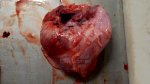
Fibrinous pericarditis - Atlas of swine pathology

Fibrinous pericarditis

Where: cardiovascular system

Possible causes: Enzootic Pneumonia (EP)Glässer diseaseStreptococcal infectionsOther

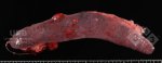
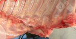
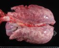
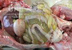
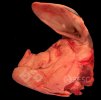
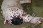
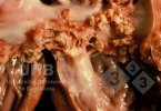
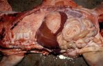
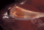
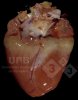
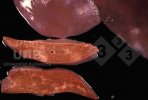
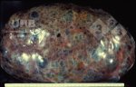
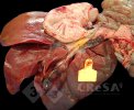
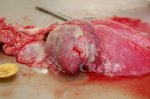
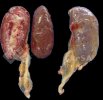
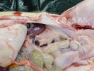
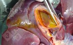
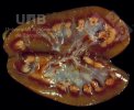
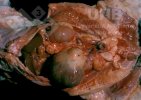
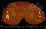
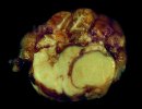
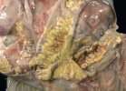
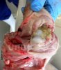
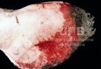
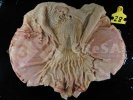
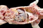
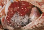
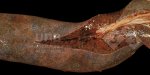
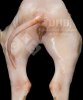
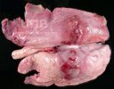
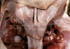
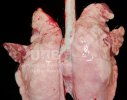
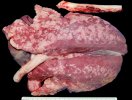
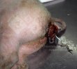
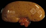
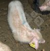
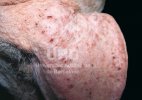
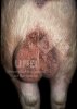
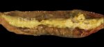
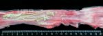
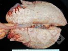
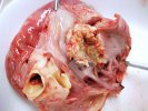
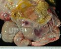
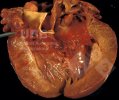
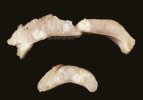
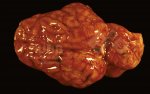
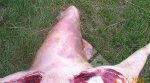
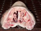
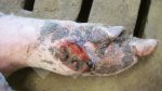
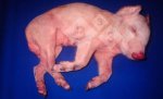
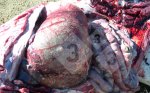
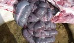
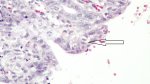
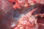
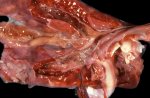
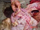
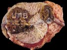
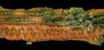
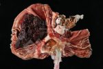
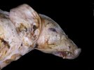
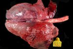
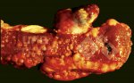
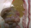
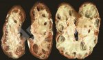
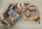
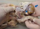
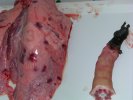
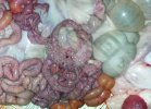
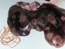
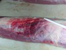
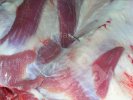
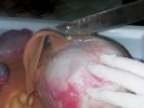
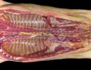
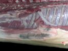
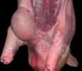
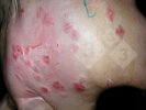
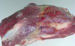
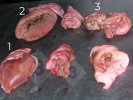
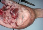
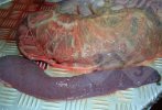
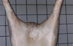
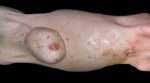
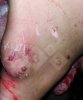
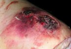
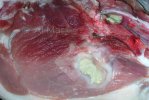
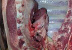
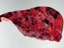
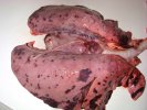
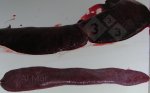
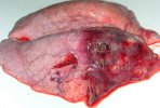
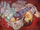
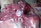
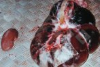
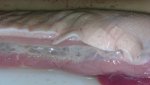
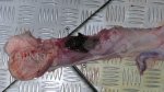
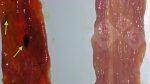
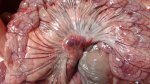
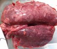
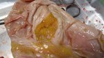
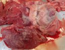
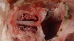
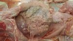
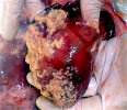
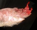
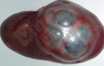
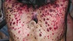
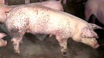
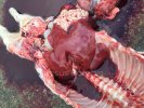
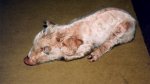
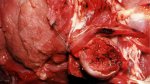
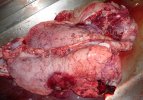
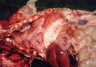
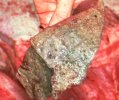
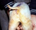
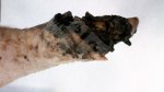
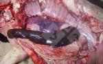
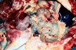
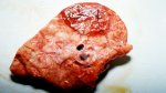
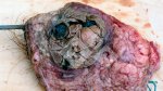
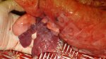
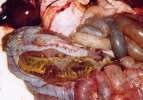
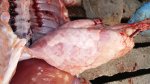
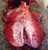
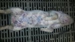
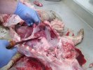
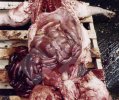
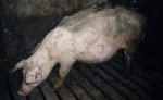
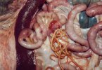
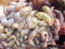
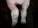
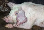
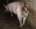
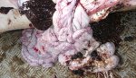
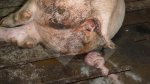
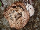
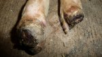
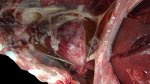
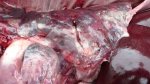
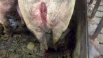
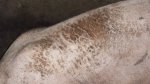
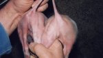
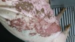
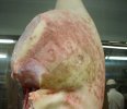
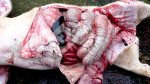
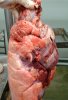
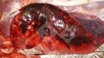
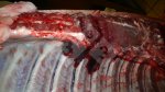
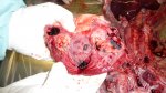
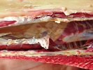
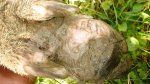
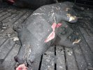
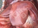
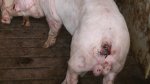
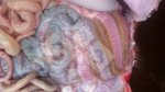
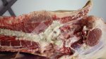
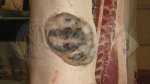
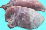
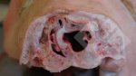
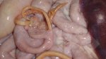
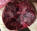
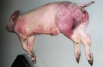
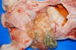
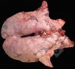
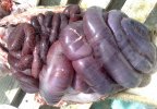
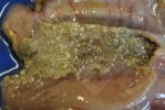
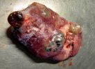
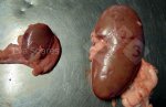
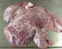
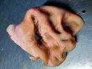
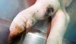
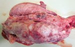
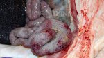
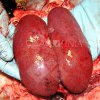
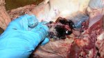
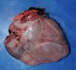
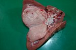
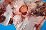
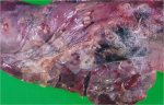
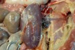
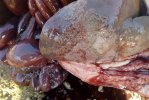
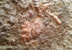
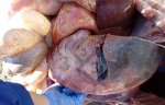
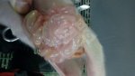
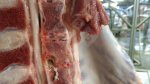
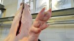
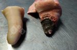
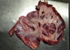
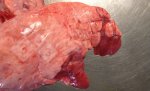
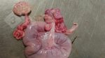
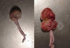
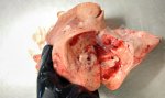
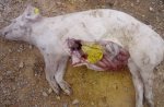
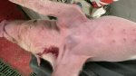
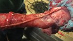
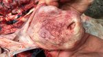
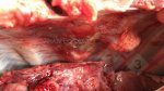
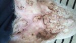
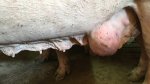
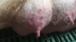
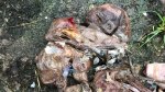
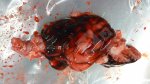
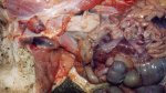
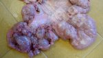
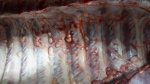
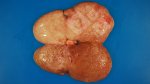
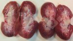
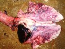
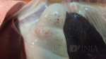
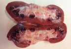
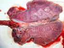
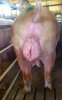
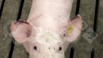
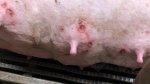
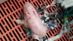
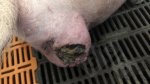
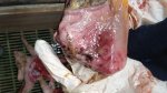
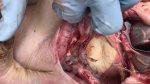
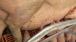
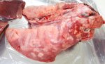
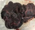
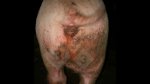
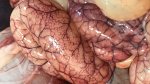
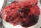
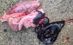
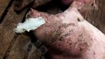
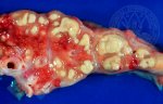
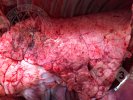
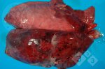
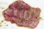
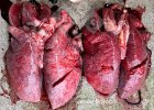
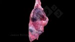
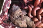
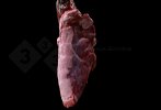
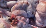
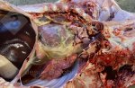
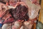
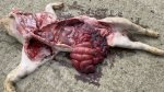
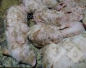
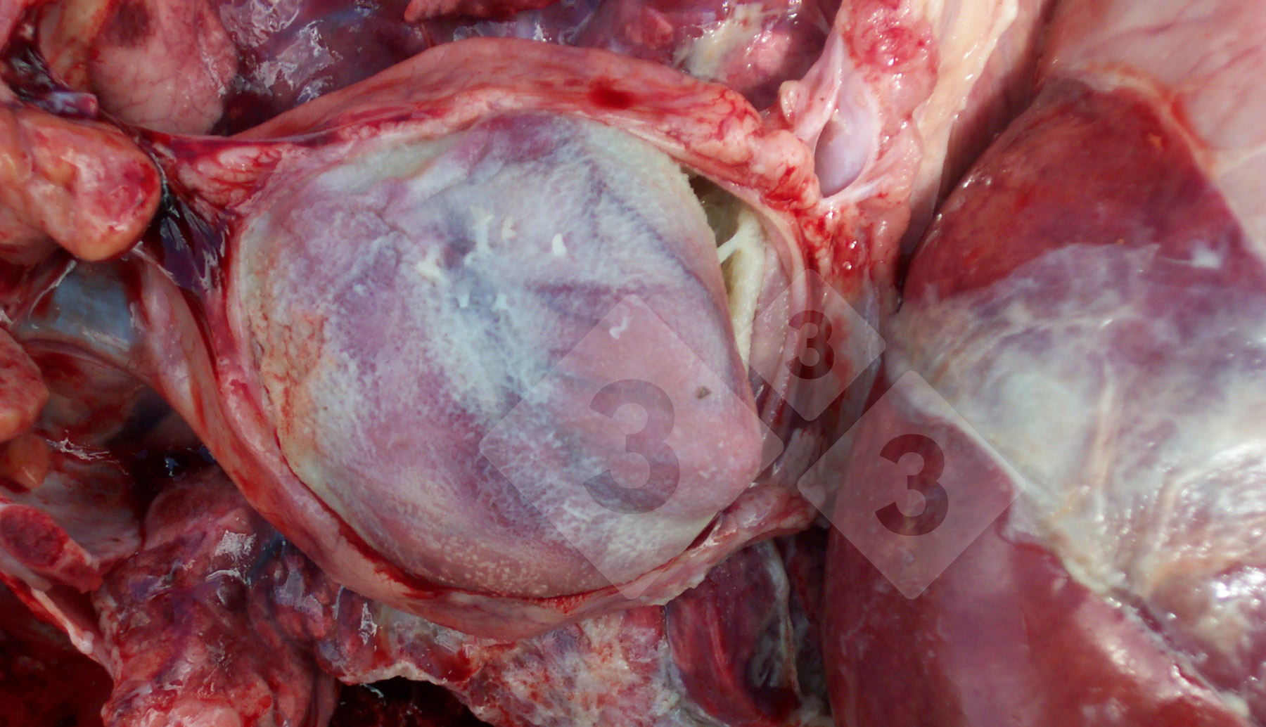
The autopsy signs are consistent with pericarditis and diffuse serositis in pigs caused by the bacterium Haemophilus parasuis. This common disease is also known as Glasser’s disease. The hospital pens of the nursery area may contain many runted, sick and dull pigs, without a clear pattern of diarrhoea or coughing. At autopsy, a dull pale thickening in patchy strands and sheets across the serosal linings of the lungs, heart and abdominal organs is noted. In severely affected pigs, this patchy grey-yellow thickening is thicker and can resemble two pieces of bread and butter stuck together. The bacteriology laboratory may have difficulty at culturing this bacterium from fresh samples. Piglets in stable H parasuis herds are usually infected within a few days of birth from their mother and develop a sub-clinical infection and acquire protective immunity. However, where piglets are mixed with piglets from other breeder pig groups, which may have different strains of H parasuis, then piglets will lack appropriate maternal antibody and develop Glasser’s disease. Use of in-feed antibiotic therapy to control this disease in weaner pigs is currently an essential part of pig raising. This form of control has the advantages of not requiring detailed knowledge of maternal antibody status, nor of the dominant strains of H parasuis present on the affected farm. Other causes of fibrinous pericarditis may include Streptococcus suis, Mycoplasma hyopneumoniae, Mycoplasma hyorhinis.

Steven McOrist
Where

cardiovascular system

digestive system

intestines

liver

mouth

stomach

genitourinary system

bladder

female genitourinay tract

kidney

male genitourinay tract

mammary gland

lymphatic system

lymph nodes

spleen

tonsils

musculoskeletal system

nervous system

other

respiratory system

lungs

nasal cavity

skin and subcutaneous tissue

Disease

Diseases caused by bacteria

App

Clostridium difficile

Clostridium novyi

Clostridium perfringens

Mycoplasma suis

Actinobacillosis

Anthrax

Atrophic Rhinitis

Bordetelosis

Brucellosis

Colibacillosis

Colitis

Edema disease

Enzootic Pneumonia (EP)

Erysipela

Exudative Epidermitis

Glässer disease

Ileitis

Leptospirosis

Mycoplasma arthritis

Pasteurellosis

Postpartum Agalactia Syndrome

Salmonellosis

Streptococcal infections

Swine dysentery

Tetanus

Tuberculosis

Yersinia infection

Diseases caused by virus

African swine fever

Aujeszky's disease

Blue eye disease

Classical Swine Fever

Encephalomyocarditis

Foot-and-mouth disease

Influenza

Japanese B Encephalitis

Nipah virus disease

Porcine circovirosis

Porcine cytomegalovirus

Porcine epidemic diarrhea

Porcine parvovirus infection

Porcine respiratory coronavirus

PRRS

Rotavirus infection

Swine pox

Swine vesicular disease

Teschen disease

Transmissible gastroenteritis

Vesicular exanthema

Diseases caused by parasites

Ascariasis

Coccidiosis

Cryptosporidiosis

Lice infestation

Mange

Metastrongylosis

Trichinellosis

Trichuriasis

Nutritional deficiencies

Biotin deficiency

Iron deficiency anemia

Mulberry heart disease

Osteoporosis, ricketts, Vit D deficiency

Toxicoses

Aflatoxicosis

Ergotism

Fumonisin toxicosis

Salt poisoning

Vomitoxicosis

Zearalenone toxicosis

Other

Atresia ani

Congenital Tremor

Epitheliogenesis imperfecta

Frostbite

Gastric ulcers

Hemorrhagic bowel syndrome

Hernias

Osteochondrosis

Other

Pityriasis rosea

Porcine stress syndrome

Rectal prolapse

Rectal stricture

Shoulder ulcers

Splay leg

Sunburn

Thrombocytopaenic purpura

Torsion of the stomach and the intestines

Uterine Prolapse

Vaginal and cervical prolapse

Vices

E-diagnostics

Pig disease diagnostic tool

access

Diseases manual

Description of the most important diseases and conditions in pigs

access