

Viruses are important causal agents of diseases affecting the respiratory system of the pig. These include porcine reproductive and respiratory syndrome virus (PRRSV), porcine circovirus type 2 (PCV2), swine influenza virus (SIV), and porcine respiratory coronavirus (PRCV). The first three are considered primary agents, capable of causing harm by themselves, while the fourth is a secondary agent, acting when there has been a previous infection by another primary agent or the animal is immunosuppressed.
PRRSV is an RNA virus, belonging to the order Nidovirales, family Arteriviridae, which is currently included in the new gen

us Betaarterivirus (formerly genus Arterivirus). Its noticeable genetic diversity has led the two genotypes of PRRSV (European and North American) to be considered as two different viral species: Betaarterivirus suid 1, corresponding to genotype 1 or European (PRRSV-1), whose prototype is the Lelystad virus (LV); and Betaarterivirus suid 2, corresponding to genotype 2 or North American (PRRSV-2), whose prototype is the VR-2332 strain.
PRRSV is able to modulate and even evade the host's immune response. This virus infects the alveolar macrophages, its main target cell, altering their function and causing their death by necrosis and/or apoptosis, which delays the onset of an effective adaptive immune response and predisposes the animal to secondary infections.
The respiratory form occurs mainly in growing and finishing pigs, and the lesions are characterized by interstitial pneumonia. Macroscopically, the lungs do not collapse when removed from the thoracic cavity, presenting a rubbery consistency and multifocal patchy areas of redness distributed throughout the lung parenchyma (Figure 1). At the microscopic level, there is a thickening of the alveolar septa associated with infiltration of lymphocytes and macrophages, as well as hyperplasia and hypertrophy of type II pneumocytes (Figure 2).
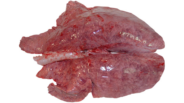
Figure1: Interstitial pneumonia caused by PRRSV.
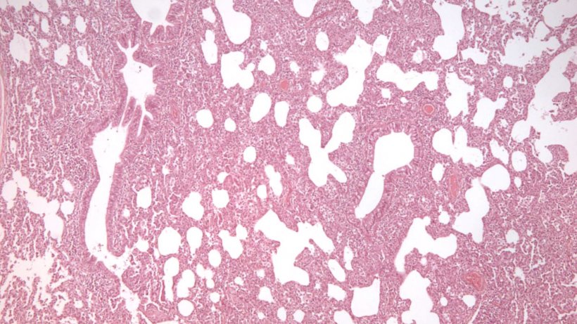
Figure2: Interstitial pneumonia, characterized by thickening of the alveolar walls, in a PRRSV infection.
PCV2- is a small, non-enveloped, single-stranded DNA virus belonging to the genus Circovirus, and the family Circoviridae. It is the causative agent of a number of diseases known as circovirus-associated diseases, including PCVAD (Porcine circovirus associated disease) - classically known as post-weaning multisystemic wasting syndrome (PMWS), PCV-2 respiratory disease, PCV-2 enteric disease, PCV-2 reproductive failure, and porcine dermatitis and nephopathy syndrome (PDNS).
PCV2 multiplies in various cells of the immune system such as macrophages, dendritic cells or lymphocytes, causing lymphoid depletion and reducing the host's ability to respond to primary and/or secondary pathogens.
The lung lesions caused by PCV2 are macro- and microscopically similar to those caused by PRRSV. Since it causes interstitial pneumonia like PRRSV, we must turn to other diagnostic techniques such as immunohistochemistry (Figure 3), in situ hybridization, or PCR in order to determine the diagnosis. It is also common for these two agents to be isolated together since PRRSV infection is associated with increased replication and virulence of PCV2.
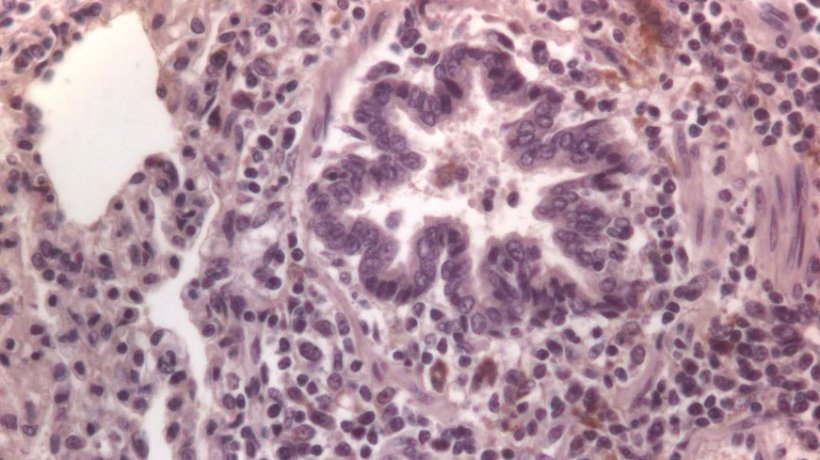
Figure 3: Cells immunolabelled for PCV2 in the lung.
SIV is a polymorphic, enveloped virus that belongs to the family Orthomyxoviridae, which includes seven different genera: Influenzavirus A, B, C and D, Isavirus, Quaranjavirus and Thogotovirus. Although the genera Influenzavirus A, B, C and D have been identified in the pig, most of the strains that cause swine flu or influenza are included under the Influenzavirus A and C genera.
SIV replicates in the epithelial cells of the upper and lower respiratory tract, altering the functioning of the mucociliary system, which in turn promotes the development of secondary bacterial infections. The main lesion it causes in the lung is bronchointerstitial pneumonia, characterized macroscopically by the presence of diffuse, dark red or brown cranioventral or multifocal lesions of variable sizes, very similar to those caused by Mycoplasma hyopneumoniae (Figure 4). At a microscopic level, it causes necrotizing bronchiolitis (Figure 5) which is accompanied by the presence of peribronchial lymphohistiocytic infiltrates in the alveolar septum and a mucopurulent exudate in the bronchioles.
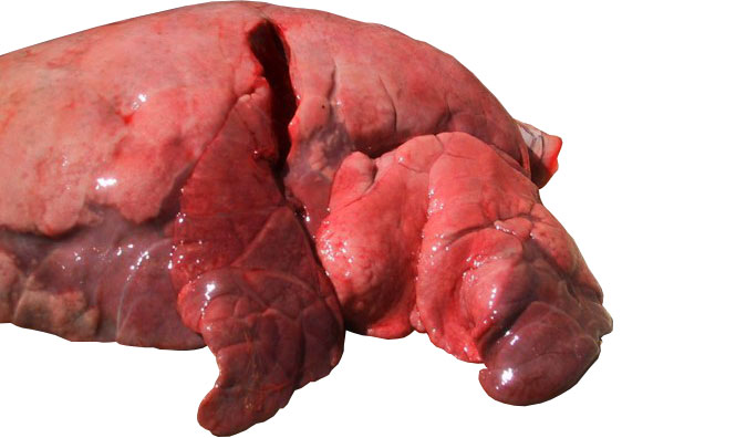
Figure 4: Cranial and middle lobes affected by bronchointerstitial pneumonia caused by SIV
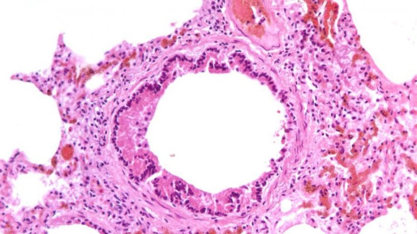
Figure 5: Necrotizing bronchiolitis caused by SIV infection
Porcine respiratory coronavirus (PRCV) is a deletion mutant of the transmissible gastroenteritis virus with a special tropism for the respiratory tract. It infects the epithelial cells of the nasal cavity, trachea, bronchi and bronchioles (Figure 6) and type I and II pneumocytes. Lung lesions are characterized macroscopically by the presence of red to brown consolidated areas with irregular borders located mainly in the middle and accessory lobes (Figure 7).
Microscopically, the lesion pattern is similar to multifocal bronchointerstitial pneumonia characterized by necrosis of the cells in the bronchial and bronchiolar epithelium, squamous metaplasia. there is also peribronchial, perivascular and alveolar septa lymphocyte infiltration, which is accompanied by the proliferation of type II pneumocytes and the presence of necrotic epithelial cells and leukocytes in the lumen of the airways and alveoli.
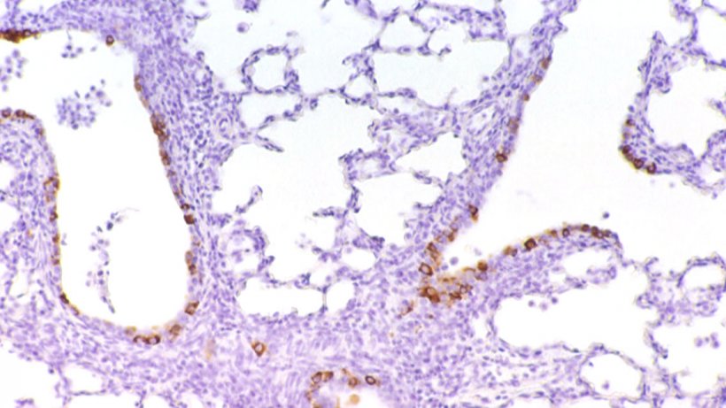
Figure 6: Cells immunolabelled for PRCV in the bronchial epithelium

Figure 7: Bronchointerstitial pneumonia caused by PRCV.
Porcine respiratory disease complex is a multietiological disease and PRRSV, PCV2, and SIV play a fundamental role, since they will cause a delay in the immune response (PRRSV, PCV2) or a failure in the functionality of the mucociliary system (SIV).


