

In the field, respiratory problems in pigs are rarely caused by a single pathogen. Usually, several pathogens are acting at the same time, which when added to inadequate management and environmental conditions, leads to the process known as porcine respiratory disease complex (PRDC).
In most cases, the main primary agents affecting the respiratory system are always involved: Porcine reproductive and respiratory syndrome virus (PRRSV), porcine circovirus type 2 (PCV2), swine influenza virus (SIV), and Mycoplasma hyopneumoniae (Figure 1), which will be accompanied by opportunistic bacteria that take advantage of the primary infection to develop their pathogenic action, such as Pasteurella multocida, Bordetella bronchiseptica or Glaesserella parasuis, among others.

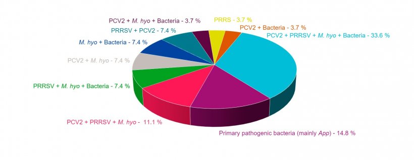
Figure 1: Combinations of pathologies found in cases of PRDC diagnosed in the last four years by the Pathological Anatomy Service of the University of Murcia's Veterinary Science Department.
In Figure 1, we can see how M. hyopneumoniae is the agent involved in most of the co-infections found in the lungs of animals diagnosed with PRDC. This pathogen, which attacks the cilia of the epithelial cells in the bronchi and bronchioles, altering the mucociliary system, also causes hyperplasia of the lymphoid tissue associated with the respiratory mucosa. Both actions facilitate the colonization, proliferation, and pathogenic mechanisms of other primary and secondary agents, that affect the respiratory system of the pig. Different experimental studies have shown that in addition to facilitate the action of other pathogens, it usually potentiates them, so that the combinations in which M. hyopneumoniae appears are usually those with the worst consequences for the affected animals, and therefore, for the farm's profitability.
One of the most frequent co-infections on pig farms with cases of PRDC is a co-infection produced by M. hyopneumoniae and PRRSV (Figure 2). In those cases where this co-infection has been diagnosed, a positive correlation has been observed between the animals that are seropositive for each of the two agents; in other words, most of the animals that are positive for PRRSV are also positive for M. hyopneumoniae. In addition, it has been proven that M. hyopneumoniae prolongs and increases the pneumonia produced by PRRSV, which could be due to the fact that in animals co-infected with both pathogens there is an increase in pro-inflammatory cytokines, such as IL1-β, IL-8 and TNF-α, which increase the inflammatory response at the lung level; but there is also an increase in the synthesis of IL-10, which is an anti-inflammatory cytokine that inhibits and modulates the production of other cytokines and would allow the damage to be perpetuated at the lung level by impeding an uncontrolled inflammatory response from being triggered. IL-10 is also capable of modulating the animal's immune response, which together with the activation of other mechanisms, alters the adaptive immune response, allowing the pathogen to continue its action in the lung for longer.
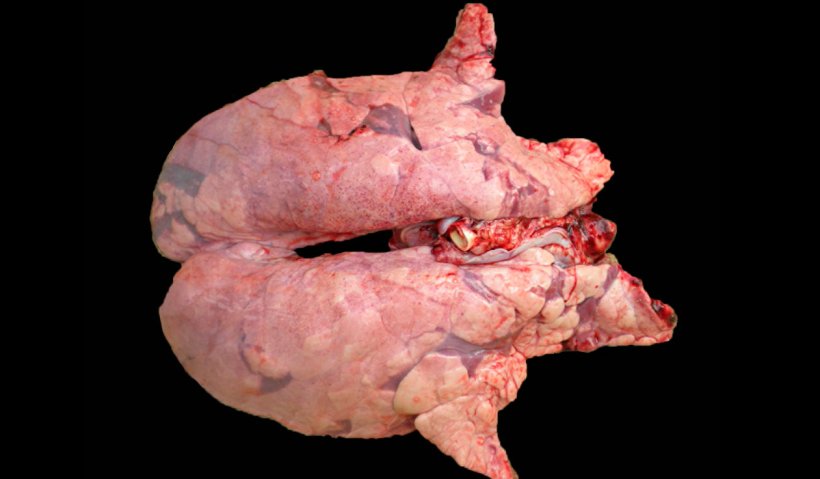
Figure 2. Pig lung co-infected with M. hyopneumoniae and PRRSV. Non -collapsed lung with reddish-brown areas of lesions distributed throughout the parenchyma, typical lesions of PRRS, which at the same time present reddish areas of consolidation in the cranioventral areas, produced by the M. hyopneumoniae infection.
In the case of co-infection with M. hyopneumoniae and PCV2, it has been shown that M. hyopneumoniae eases the severity of the lesions produced by PCV2 in the lung and lymphoid organs, promoting greater virus replication, as well as its persistence in the tissues (Figure 3), thus increasing the incidence of cases of PCV2-systemic disease (actual name given to tPMWS (postweaning multisystemic wasting syndrome).
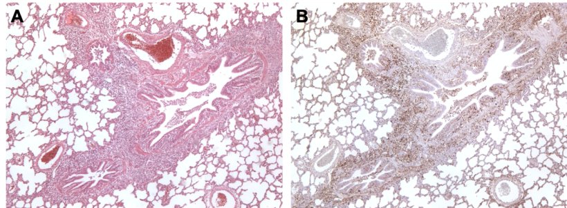
Figure 3. Pig lung co-infected by M. hyopneumoniae and PCV2. A: Area of peribronchiolar lymphoid hyperplasia caused by M. hyopneumoniae. B: Large amount of PCV2 antigen in this same area of lymphoid hyperplasia.
In animals with co-infections of M. hyopneumoniae and SIV (Figure 4) it has been observed that when pigs are infected with a low virulence strain of swine influenza virus after infection with M. hyopneumoniae there is a significant increase in clinical signs and lung lesions, suggesting that mycoplasma is capable of increasing the virulence of these SIV strains.
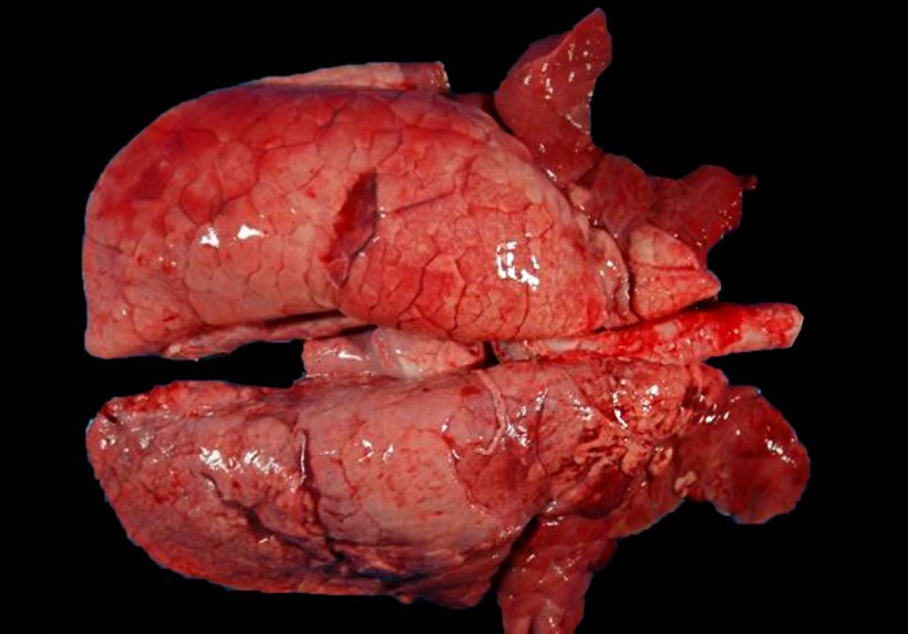
Figure 4. Pig lung with co-infection of M. hyopneumoniae and SIV. Red areas of consolidation in the cranioventral portions of the lung and some smaller areas in the diaphragmatic lobe. In these craniovental lesions, there are both lesions from M. hyopneumoniae and SIV, making it imposible to differentiate between them macroscopically.
M. hyopneumoniae also appears frequently associated with other bacteria, whether they are primary agents, such as Actinobacillus pleuropneumoniae (Figure 5), or opportunistic agents, such as Pasteurella multocida (Figure 6), enhancing their action in the lung.
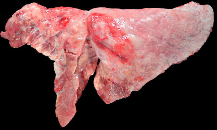
Figure 5: Pig lung co-infected with M. hyopneumoniae and A. pleuropneumoniae. Cranioventral areas of consolidation with a reddish-brown color, caused by infection by M. hyopneumoniae, and an oval-shaped lesion with pleural fibrosis and hemorrhagic area in the diaphragmatic lobe, which would correspond to the chronical stage after an outbreak of necrosis caused by A. pleuropneumoniae.
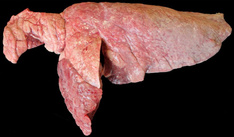
Figure 6. Pig lung co-infected with M. hyopneumoniae and P. multocida. Areas of reddish-brown cranioventral consolidation, elevated with respect to the rest of the lung parenchyma, which would be consistent with a lesion caused by P. multocida. In the cranial lobe and cranioventral margin of the caudal lobe there are reddish depressed areas of consolidation which correspond to lesions caused by M. hyopneumoniae.
As we have seen, when M. hyopneumoniae appears in co-infections together with other primary agents of PRDC, it acts boosing and prolonging the lesions caused by these pathogens, which is why its control is crucial to minimize the effects of PRDC and reduce the economic losses associated with the disease.


