General description of the situation
This clinical case began in the summer of 2023 and affected numerous finishing farms, mainly in the Spanish areas of Castilla-Leon and Castilla-La Mancha.
Most cases were associated with animals with a high feed intake capacity, affecting a proportionally higher number of Duroc or Iberian genetic crosses.

The clinical cases were not related to specific sow farms or production companies.
Clinical symptoms and necropsies
In September 2023, several affected finishing barns were visited and the clinical symptoms and necropsy findings were as follows:
- Pale, prostrate, listless pigs with no fever
- Very high mortality in the final stages of finishing, from 70 kg live weight onwards. In some cases, the mortality rate increased greatly, reaching 20-30% and even up to 40%.
- Dark-colored urine (Video 1).
- Liver damage, jaundice (Photos 1 and 2).
- Hemorrhagic gastroesophageal ulcer (Photo 3).

Photo 1. Marked jaundice in an affected pig.
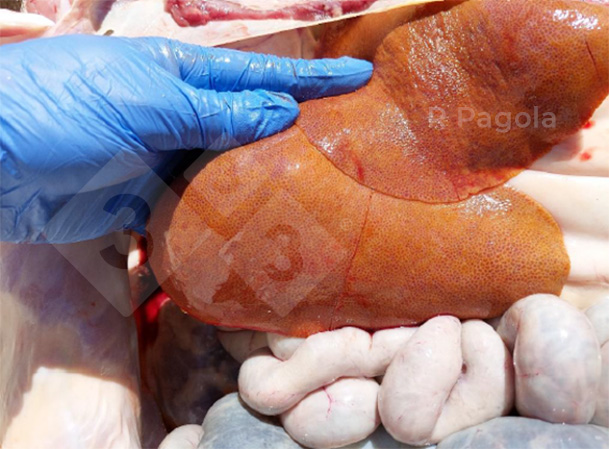
Photo 2. Appearance of a liver from the necropsy of an affected pig.
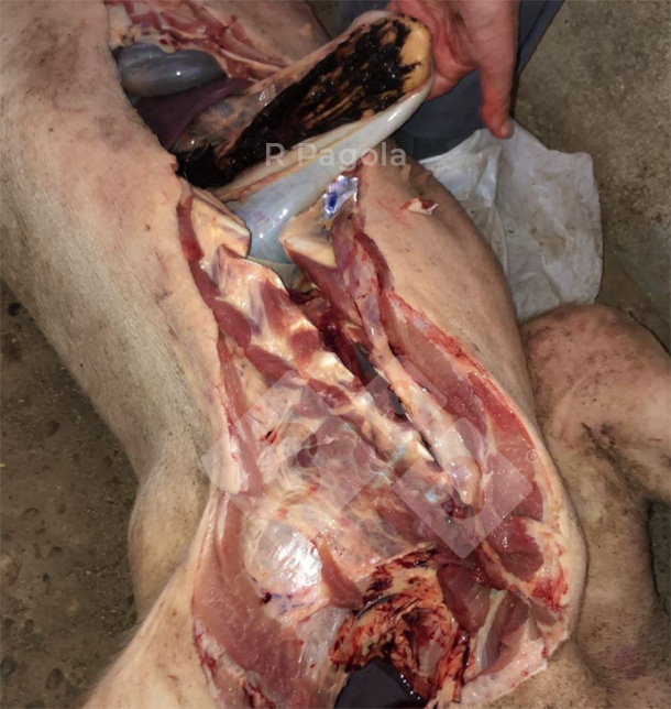
Photo 3. Hemorrhagic gastric ulcer of an affected pig.
Although initially the focus of the symptoms was on the ulcers, after the first visits it seemed clear that the ulcers were a secondary effect related to the main problem. The liver damage was attributed more importance and poisoning was suspected.
Video 1. Dark-colored urine from an affected pig.
Health status of the animals
A differential diagnosis was made for infectious agents such as PRRS, APP, swine dysentery, ileitis, and PCV-2, and there was no evidence that the problem was associated with any of these pathogens. More than twenty finishing barns with this problem were visited in both affected regions. Many of them were negative for these pathologies.
Blood tests
The blood of animals with clinical symptoms revealed a vitamin E deficiency, as shown in Table 1, in addition to pronounced anemia, which is logical due to the hemorrhagic gastric ulcers.
Table 1. Description of vitamin E blood values in affected pigs.
| Sample reference | Vitamin E (mg/dL) |
|---|---|
| 1 Barn 1 (#1) | 1.20 |
| 2 Barn 2 (#7) | 1.70 |
| 3 Barn 2 (#8) | 0.60 |
| 4 Barn 3 (#9) Problem | < 0.4 |
| 5 Barn 3 (#10) Problem | < 0.4 |
| 6 Barn 3 (#11) | * |
| 7 Barn 3 (#12) | * |
| Normal values | 1.60 - 4.6 |
It was ruled out that the vitamin E deficiency was of dietary origin since the correct levels were found in the feed supplied.
Anatomopathological analysis
Histopathological analyses were performed on different organ samples (heart, lung, mesenteric lymph node, diaphragm, loin muscle, limb muscle, brain, spleen, liver, kidney, ileum) collected from different necropsies of pigs with clinical symptoms. Although all the organs submitted had a yellowish-orange tinge due to jaundice, the main lesions were observed in the liver samples, and the first macroscopic assessments were confirmed. Likewise, microscopic lesion analysis of the liver samples confirmed the initial macroscopic assessments:
- The liver fragments presented a yellowish coloration and a very marked lobular pattern.
- There was a very marked alteration of the hepatic lobules due to the collapse of the hepatic sinusoids and deposition of abundant mature fibrous tissue in the interstitium, which was more pronounced in the periportal region.
The few remaining hepatocytes showed the following changes:
- marked increase in cytoplasmic size (megalocytosis)
- presence of granular and/or ochraceous pigmented cytoplasm
- marked increase in nuclear size (megalokaryosis)
- presence of abundant nuclear inclusions with cytoplasmic-like dye appetence (pseudoinclusions)
- marked distension of the biliary canaliculi due to the accumulation of ochraceous material (canalicular cholestasis)
In the periportal area, a very marked ductular proliferation was observed. Multifocally there were scarce mononuclear infiltrates with the occasional presence of neutrophils.
The final summary of the anatomopathological diagnosis indicates:
- Severe diffuse hepatocellular degeneration with periportal to centrolobulillar fibrosis, megalocytosis, megakaryosis, and biliary proliferation.
- Coronary vasculopathy with petechial hemorrhages; multifocal, moderate, which may be associated with hyperammonemia.
- Hepatic encephalopathy, probably due to toxic damage of ammonium in the central nervous system by hepatic metabolization failure.
Toxicological analysis
Numerous toxins/pesticides/heavy metals were analyzed in the liver, as it is the target organ, but the results were inconsistent in concluding the cause of the problem.
Intense sampling was done to test for the presence of several mycotoxins in both the raw materials and the final feed, focusing on aflatoxins and fumonisins, with no clear results of contamination associated with clinical cases.
In several discussions with colleagues from other regions, we checked to ensure they were not reporting similar cases in other major pork-producing regions such as Aragon, Catalonia, and Murcia; but, they get their raw feed materials mainly from imports.
The hypothesis of poisoning was still sustained, which was the main avenue of investigation. In December it was decided to analyze the feed and cereals used to manufacture the batches of feed that the animals consumed in September and October. It should be taken into account that this problem was observed in cereal-producing regions, so the national cereal makes up a significant part of the diet, unlike in other areas of the peninsula where its use is more limited.
We analyzed pyrrolizidine alkaloids (PA) and the following results were obtained, which now help us to focus the diagnosis of the problem:
- PA levels in grains incorporated in the feed during the problematic months
- PA levels in the feed supplied during the problematic months
Results of the analyses conducted on the feed and the cereals included in the feed supplied to the pigs when clinical problems occurred showed abnormally high total pyrrolizidine alkaloid values. In the case of the grain analyzed, the results show PA values around 12,000 ppb. For the feed supplied during the problematic months, the values are around 4,000 ppb, as shown in Table 2.
Table 2. Average total PA values in grains and feed associated with cases with affected animals.
| Grain | Feed | |
|---|---|---|
| Total pyrrolizidine alkaloids (μg/kg) | 12,530 | 4,285 |
The EFSA (European Food Safety Authority) determines the amount of PA that would become toxic if consumed for six months to be 15 µg/kg of live weight/day.
Considering these quantities, the calculation for a pig weighing about 70 kg would be 1050 µg of PA per day as a toxic limit. If we estimate consumption at 2 kg of feed per day with 4000 µg of PA in the feed, it would give us the amount ingested by the animal to be 8000 µg/day which is 7.5 times above the toxic limit determined by the EFSA.
What are pyrrolizidine alkaloids?
PA are natural toxins, products of the secondary metabolism of weeds such as Senecio Vulgaris, Crotolaria, Heliotropum, etc. The possible cause of PA appearing in the grains used in the manufacture of animal feed may be due to the poor quality of the 2023 harvest, as a consequence of the drought suffered in the central zone, and perhaps the reduced use of adequate herbicides in the grain fields.
PA have a common toxicity profile, resulting in varying degrees of progressive liver damage (centrolobular hepatocellular necrosis) and veno-occlusive disease. All this would explain the symptoms we saw in the field (hepatic alterations and gastric ulcers). They can also cause pulmonary toxicity.
The low vitamin E concentration in the blood of affected animals indicates oxidative stress of the animal as a consequence of hepatic degeneration. It is well-referenced that such hypovitaminosis E is associated with the development of gastric ulcers.
Measures taken
- Limited use of national grains in all feeds.
- Requested feed and national grain analyses to monitor the presence and concentration of PA.
- Provided support with liver protectants until the risk was eliminated, sequestrants with proven efficacy for these alkaloids, and products with buffering capacity to control the pH in the stomach when ulcers are observed.
Evolution of the problem
Once the root cause of the problem (pyrrolizidine alkaloids) had been diagnosed and the corresponding preventive measures were taken, we saw the levels of PA decreased by 98%, and consequently, the clinical symptoms that had been observed months prior progressively disappeared.
Conclusions
Based on the tests and analyses performed, we can conclude that the definitive diagnostic cause was toxic concentrations of pyrrolizidine alkaloids from some locally harvested grain fields, which led to liver damage, decreased levels of vitamin E in the blood, and gastric ulcers, resulting in the death of a significant number of affected pigs.