Introduction
An essential component in the control and elimination of Mycoplasma hyopneumoniae (M. hyo) is the assessment of the duration of shedding of the bacteria in swine populations, since that will determine the timing of introduction of replacement females, the duration of herd closure. Most importantly, such information will allow the development of more appropriate gilt acclimation strategies. Although the duration of shedding in experimentally infected pigs has been assessed, the epidemiology of M. hyo in naturally infected gilt populations remains largely unknown (Pieters et al, 2009; Pieters et al, in press). The objective of this study was to describe the pattern of M. hyo infection and persistence in a gilt population naturally exposed.

Materials and methods
The farm enrolled in this study had recently experienced a M. hyo outbreak, after being negative for over 5 years. In this prospective cohort study a group of 63 gilts were randomly selected at 21 days of age, tagged and sampled via laryngeal swabbing weekly for 5 weeks and monthly afterwards. Serum samples were collected at 21, 110 and 140 days of age. A final sampling occurred during the farrowing stage, where a tracheal sample was collected from study sows and 5 of their piglets. Troubled by the poor sensitivity of the laryngeal method in mid to late infection, it was decided to adopt tracheal sample collection instead, starting at 200 days of age (Fablet et al, 2010). Tracheal samples were collected by inserting the end of a post cervical artificial insemination (PCAI) rod into the trachea. After sample collection, the connection piece was then detached from the rod and inserted into a sterile snap cap tube with 0.5 mL of PBS and refrigerated at 4°C. With the objective of assessing infection in the lung, a total of 20 study gilts were humanely euthanized and subjected to a comprehensive pathological examination. All laryngeal, tracheal and lung samples were submitted to the Veterinary Diagnostic Laboratory at Iowa State University and tested by quantitative M. hyopneumoniae PCR, serum samples were tested for M. hyo antibodies by IDEXX ELISA.
Preliminary results and discussion
Over the course of the study, 19 gilts either died or were discarded at different stages of the study due to them not meeting the gilt selection criteria (Figure 1). At weaning, 11% of the gilts were infected and the last positive gilt was detected positive at 284 days later. Despite the recent M. hyopneumoniae outbreak the prevalence at weaning was low. It is hypothesized that the lower prevalence could be due to the administration of in-feed antibiotic in the sows, a less virulent M. hyo strain or the lack of sensitivity of the laryngeal method for early detection. The prevalence at 80, 110, 140 and 170 days of age was 14.5%, 29.7%, 28.8% and 4.2%, respectively. This slow transmission and low exposure level, even under an outbreak scenario, represents one of the biggest challenges for M. hyo control.
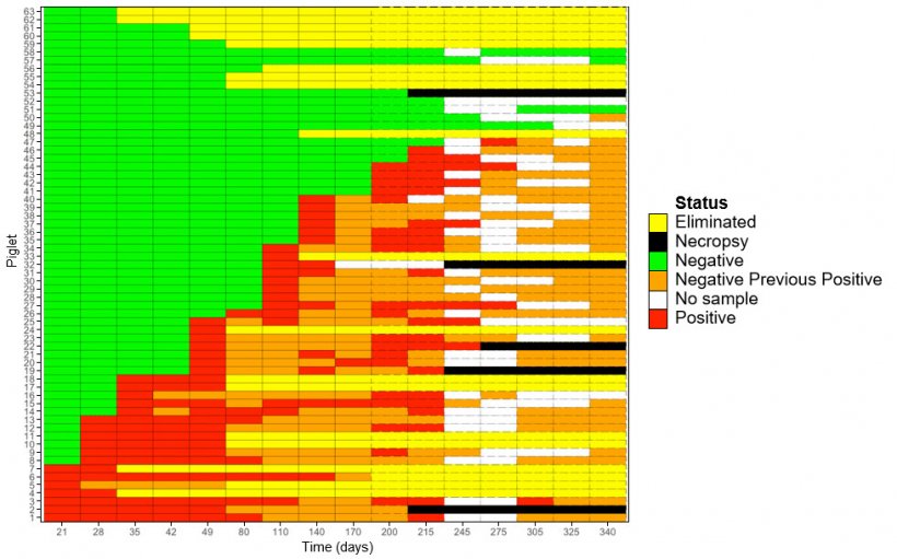
The early decline in detection led to the decision to utilize a more invasive and sensitive diagnostic method. A side by side comparison was performed using laryngeal swabs and tracheal samples. The detection of M. hyo in laryngeal swabs was 8.8% compared to 42.2% in tracheal samples at 200 days of age. At 215 days, 5.2% of laryngeal swabs tested PCR positive compared to 52% using tracheal samples. The latter sampling method appears to be a more sensitive methodology for detection of M. hyo during chronic infection (Pieters et al, in press; Fablet et al., 2010). The prevalence decreased to 33.3%, 18.75%, 5.80%, 0% and 0% at 245, 275, 305, 325 and 340, respectively. In this study 4 gilts (6.3%) remained PCR negative throughout the study; however; there was evidence of seroconversion in 100% of the gilts by 140 days. This reveals that although, 93.4% of gilts in this study showed at least one PCR positive laryngeal or tracheal sample, not all exposed gilts were detected positive, even after consecutive samplings. This limitation of sensitivity should be taken into account when designing sampling strategies for M. hyo (i.e. determining day “zero” or detection prior to reopening a herd). Finally, at farrowing 20 study sows and 5 of their due-to-wean piglets all tested PCR negative.
The seroprevalence at 21 days of age was 100%, evidencing a consistent transfer of maternal antibodies from sow to piglet. The seroprevalence at 110 days was 59.5%. From these seropositive gilts, 39% were PCR negative using laryngeal swabs. Seroconversion is usually seen between 4-8 weeks after infection, consequently these results would indicate that the laryngeal swabbing method was not effective for detection of infection in previous samplings. In contrast, 38% of the gilts were serologically negative at 110 days of age, from these, 27% had previously tested PCR positive by laryngeal swabs between 21 and 49 days of age. This shows the variability in seroconversion for M. hyo and supports the fact that antibody response is a lagging indicator for exposure. The seroprevalence at 140 days revealed that 100% of the gilts had seroconverted, still 30% of seropositive gilts were not detected PCR positive by laryngeal swabs after 8 different sampling events.
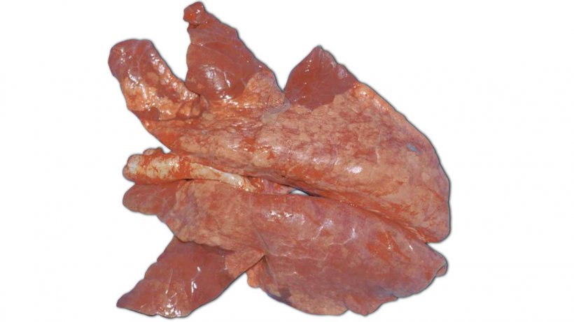
Histopathology performed on the lungs of 2 gilts necropsied at 215 days of age revealed lymphocytic peribronchiolitis, congestion, with previous bronchopneumonia of unknown etiology (Foto 1). All lung sections and tracheal samples tested PCR negative for M. hyo. At 245 and 275 days, lung samples from 3 gilts showed no gross or histopathological evidence of M. hyo disease and all samples tested PCR negative. However, the tracheal samples from these gilts collected prior to euthanasia tested PCR positive. The latter observation could indicate a potentially lower sensitivity of bronchial samples collected for detection of M. hyo compared to tracheal samples during chronic infection.
Take-home messages
- Shedding and exposure of piglets to M. hyo in the farrowing room, even under recent introduction into a negative herd, can be low.
- The peak of acute infection using laryngeal swabs was observed at 49 days of age (39%). The peak of chronic infection was observed at 215 days (52%).
- M. hyo transmits slowly within exposed populations. Even during outbreak events, the proportion of positive gilts at one time never exceeded 52%.
- The last positive gilt was detected at 284 days. This provides further evidence that populations can shed for long periods of time.
- Laryngeal swabs appear to lack sensitivity for M. hyo detection in mid to late infection, as evidenced by seroconversion coupled with negative laryngeal swabs by PCR.
- Tracheal samples appear to be more sensitive diagnostic tool compared to laryngeal swabs.
- M. hyo antibody response is a lagging indicator for exposure.