Introduction
Inaba et al. (1983) were some of the first authors to demonstrate the utility of B-mode ecography in sow pregnancy check. In the following years, several studies were conducted. In these studies the superiority of ultrasonography was repeatedly demonstrated over other diagnostic methods such as the observation of returns in heat and the endocrinological tests.

More recent works have confirmed the validity of the ultrasound method also for the visualization of ovarian structures in the peri-ovulatory period and in the diagnosis of ovarian pathologies.
Ultrasound basis, materials and use
There are different types of probes on the market which differ in shape, frequency and type of use: in veterinary medicine, mainly linear probes and sectorial probes are used.
The greatest versatility for the observation of the sow's reproductive tract is given by a frequency of 5MHz; in various works, however, other frequencies (3.5 Mhz and 7.5Mhz) have been used for the diagnosis of pregnancy and the examination of the ovary.
Classic pregnancy check vs early pregnancy check
A probe with a frequency of 5MHz can determine pregnancy starting from day 20-21, while with an ultrasound machine with a 7.5MHz probe or a good ultrasound system with an electronic probe, pregnancy can be detected as early as 18 days.
The early diagnosis of pregnancy with a 7.5MHz probe has the main great advantage of identifying non-pregnant sows and, together with the assessment of the ovarian status, allows the timely reintroduction of the returning sows into production, avoiding the accumulation of non-productive days; it also allows the individual assessment of the uro-genital tract to search for any physio-pathological abnormalities, accelerating decisions regarding treatment or any culling.
Ultrasound puberty check
It has been shown that the use of ultrasound in the evaluation of the uterus and ovaries is a valid tool for diagnosing the state of puberty of gilts.
The exclusive presence of small follicles (2-5 mm of diameter) in the absence of corpora lutea is a typical finding of pre-pubescent gilts. The size of the uterus can also be useful in determining the state of sexual maturation of the reproductive system: Martinat Botté et al. (2003), were the first to evaluate the size of the uterus, defining "uterine area" as the area that includes all visible parts of the uterus within the ultrasound image.

Kauffold et al. (2004) also used the size of the uterus to assess the state of puberty by measuring a cross-sectional area of the uterus in their maximum and minimum diameter.
According to these parameters, gilts that have a cross-sectional area ≤ 1cm2 are considered prepubertal, conversely, gilts that have a cross-sectional area ≥1.2 cm2 are considered pubertal.
Ultrasound staging of the ovarian cycle
At the onset of heat and up to 24 hours before ovulation, the follicles show a diameter between 7 and 9 mm, although there is a strong individual variability (Soede et al., 1998).
Close to ovulation, the pre-ovulatory follicles change their shape from spherical to ovoid / polygonal. The ovulation process can take several hours and pre-ovulatory follicles can be observed in conjunction with hemorrhagic bodies; the latter are clearly identifiable by their echogenic aspect. Ovulation is considered complete if sequential ultrasound investigations show the disappearance of all pre-ovulatory follicles or a marked reduction in the number of previously observed pre-ovulatory follicles.
Infertility and pathological cases
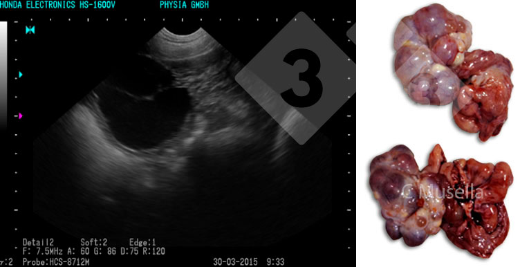
The diagnosis of single or multiple ovarian cysts is relatively simple even if the complete identification of the type of cyst through the ultrasound image is not always certain.
As for the uterine structures, the presence of fluid, not due to pregnancy, semen or estrus, must be considered pathological and indicative of an exudative inflammation of the uterus. Observation of the exudate through ultrasound is only possible if the inflammation is acute.
On the contrary, chronic endometritis, which is the most common type of inflammation in the sow, cannot be diagnosed with certainty by ultrasound.
Conclusions
In recent years, the use of ultrasound has been strongly implemented in pig farms; despite this, currently the main purpose and use of the ultrasound is still limited to pregnancy check starting from 21 days post-AI.
On the other hand, with a high-precision ultrasound system it is possible to go into details with the state of the reproductive system, investigating in particular the status of the ovaries and uterus and provide valid support to the “on farm” veterinarian and the breeder in the diagnosis of reproductive problems and in the improvement of the farm performance.