

B-Mode ultrasonography is a widely used diagnostic modality in male reproduction, particularly in men. It has been proven feasible to visualize the testes, epididymides, and the accessory sex glands when determining causes of subfertility/infertility and diagnosing diseases of the male genital tract. In veterinary medicine, ultrasonography has been used in stallion, bulls, bucks as well as dogs, but also, albeit less often, in boars, which will be shortly described in this paper.
B-Mode ultrasonography of the boar’s reproductive tract has to be performed while the boar is in an upright position and ideally also immobilized. This can be achieved by crating the boar or scanning while the boar mounts a semen collection dummy. Another approach for immobilization is to lift the boar by using special purposely-designed chutes, but this may have the limitation of a slight anatomical dislocation of the testes and epididymides. In some cases it is also possible to scan calm boars post-semen collection in the semen collection room or while being confined in their pens.

There are many ultrasound units that can be used for scanning the boar’s reproductive tract. While it is not the intention to prioritize units in this paper, the authors have gained experience in this diagnostic technique using devises which provide for high resolution including the HONDA 1600, Fazone CB and Zonare Z-one with great success.
Scanning of the accessory sex glands has to be done transrectally using a linear probe (Clark & Althouse, 2002; Fig. 1), while for the testis, epididymis as well as the spermatic cord a sector or (micro-) convex probe is the preferred device (Fig. 2A & B). Frequencies of 5.0 – 9.0 MHz have been used, with the lower frequencies being more appropriate for tissues that require a higher penetration depth (i.e. the testis and the epididymal corpus), and the higher frequencies for rather tiny structures such as the epididymal head and tail. It thus depends on the objective of the examination which will be the optimum probe and frequency.

Fig. 1: Placement of transducer holder with the transducer per rectum for visualization of accessory sex glands of the boar (from Clark & Althouse, 2002).
Prior to scanning of the testes, epididymides and spermatic cord, the scrotal surface should be cleaned and, if necessary, shaved.
The testes can be scanned either longitudinally or transversally (Fig 2A & B). To determine the testis’s circumference, transversal imaging has to be employed. Healthy testicular tissue has a medium echogenicity and is of homogenous echotexture, with the hyperechogenic rete testis in the testis center (Fig 3A & B).

Fig. 2: Procedure of scanning of the testis in a non-restrained calm boar post-ejaculation in the semen collection room. A) Longitudinal scanning using a micro-convex probe. B) Transversal scanning using a convex probe. This scanning location has also to be employed when imaging the epididymal corpus.
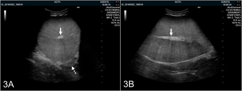
Fig. 3: Ultrasonographic images of the testis of a boar scanned transversally (A) and longitudinally (B). The rete testis is imaged as a hyperechogenic spot in the testis’s center (solid arrow). The testicular parenchyma appears medium echogenic and of homogenous echotexture. Transversal imaging also provides optimal visualization of the epididymal corpus (dotted arrow), which is normally of similar ultrasonographic appearance as the testis.
Scanning of the epididymis has to be done at various positions depending upon whether the head (from ventral), corpus (transversally in the middle of the horizontal axis of the testis while manually fixing the testis) or tail (from dorsal while manually pushing the testis and epididymis dorsally) is of interest (Fig. 4; Kauffold et al., 2011).

Fig. 4: Schematic illustration of the topography of the testis and epididymis in boars, with suggested transducer placement when assessing the epididymal caput (= head; a), corpus (b) and cauda (= tail; c). The caput and cauda are best imaged longitudinally and the corpus transversally (from Kauffold et al., 2011).
Generally, the epididymal tissue is also of homogeneous and regular echotexture, with the head and corpus having a more fine and the tail a rather marbled echotexture (Fig. 5A–C). The echogenicity, however, as determined by “grey-scale-analysis” and given as the mean grey value, slightly differs between the three segments, along with changes when compared images obtained pre- and post-semen collection (Kauffold et al., 2011) from a boar.

The healthy accessory sex glands (e.g., bulbourethral, prostate and vesicular gland) have been best detailed by Clark & Althouse (2002) as follows: “The paired bulbourethral glands are best described as a long oval gland with uniformly echogenic appearance with a large anechoic space in the center of the gland extending most of its length. The walls of the vesicular glands were found to be thin, with the parenchyma having multiple small echolucent areas that appeared to merge and form a central canal. The prostate gland was best identified as a walnut-sized gland with a uniform echogenic appearance”. As has been shown for the epididymis, the architecture of all three glands changed post- if compared to pre-ejaculation in so far as the “fluid component” decreased and the glands then appear with a higher echogenicity.
Studies on the practical use of ultrasonography in boar reproduction are sparse. The diameter of the testes has been determined in order to relate to average ejaculate total sperm number (Clark et al., 2003) or to assess pubertal development (Ford & Wise, 2010). While the correlation of paired testicular diameter and total sperm number was poor, at least in boars > 8 months, the determination of the testicular size of younger boars, i.e. at approximately 4 months, seemed useful for the prediction of the testicular volume and thus daily sperm production at an older age when the boar is mature. There is a recent report on an infertile Large White boar with a multicystic degeneration of the bulbourethral gland; the affected gland was imaged with solid and well-defined anechogenic cysts of different size (Fig. 6; Grahofer et al., 2016).

Fig. 6: Images of the bulbourethral gland of the affected boar having multiple well-defined anechogenic cysts of different size (A) and of a healthy boar for comparison (B) (from Grahofer et al., 2016).
To summarize, ultrasonography of the boar’s reproductive tract is a viable diagnostic modality which can deliver valuable information with respect to tissue function and health. While practical use is currently limited, it is the author’s strong expectation that the use of this modality (in addition to others such as Color-Doopler [Fig. 7A & B]) will gain further use by the swine practitioner as a tool to predict potential ejaculate yield and to definitely diagnose subfertility/infertility associated with diseased tissues.

Fig. 7: Testis images obtained using Color-Doppler ultrasonography. A) Vessels of the spermatic cord. B) Arteria testicularis
Acknowledgement
The authors thank Nadja Legler for her help with generating images used in this article.


