Lung lesion evaluation is, by far, the information most frequently collected, basically to confirm and quantify respiratory problems, as well as to assess the outcome of certain intervention strategies. The two most prevalent pulmonary lesions observed at the pig slaughterhouse are:
- Craneo-ventral pulmonary consolidation (CVPC)
- Pleuritis, mainly in caudal lobes.
In the present article, CVPC refers to purple-dark and well-defined areas of pulmonary consolidation, mainly located bilaterally in apical, intermediate, accessory as well as the cranial part of the diaphragmatic lung lobes (Figure 1).
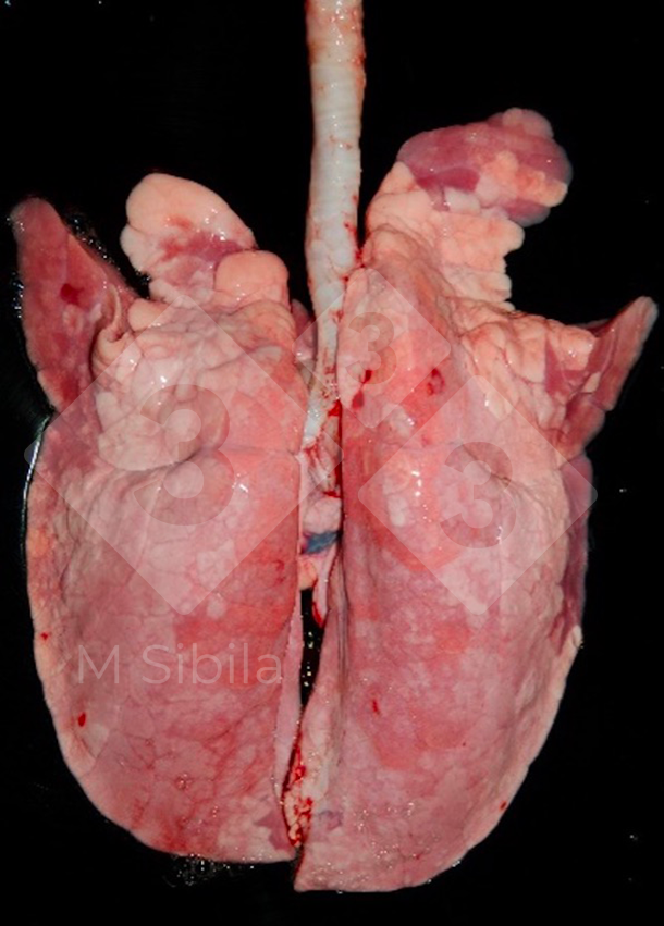

In most cases, this type of lesion is associated to Mycoplasma hyopneumoniae (M. hyopneumoniae) infection. However, this lesion can be produced by other pathogens, such as swine influenza virus in a multifocal pattern, and Bordetella bronchiseptica and Pasteurella multocida in a more diffuse manner. Therefore, the causative agent(s) should be determined by means of laboratory techniques.
In acute M. hyopneumoniae disease cases (and without secondary infections), the lesion is clear and well-demarcated, being easily recognized and scored. However, in most cases, the lesion is aggravated with the presence of other bacterial or viral pathogens, varying in color, consistency, and extension (but not the craneo-ventral location).
At slaughterhouse, CVPC lesions tend to be chronic, the color tends to be greyish, and the tissue can be retracted forming scars or interlobular fissures, complicating its observation and scoring. If other lesions are present, such as pleuritis, the scoring from the affected lobes can even be more difficult or not possible to assess. Macroscopically the severity of CVPC is measured by its extension; the higher the percentage of affected tissue, the more severe the lesion.
Pleuritis refers to the inflammation of the pleural serosa. When this lesion is confined to dorso-caudal lobes, it is strongly suggestive of Actinobacillus pleuropneumoniae infection (Figure 2). Chronic cases (those usually present at slaughterhouse) are characterized by fibrous adhesions to visceral and parietal pleura. In such cases, lung tissue adhesions are common and leave part of the organ adhered to the thoracic wall during the lung’s removal from carcass, leading to its condemnation. In this scenario, presence of these lung adherences would be indicative of high severity.
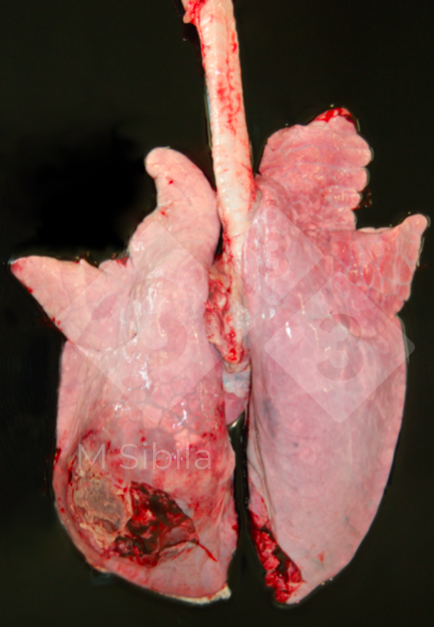
Lung lesion scoring systems are easy and non-invasive methods that provide information on prevalence and extension (but not incidence) in a relatively inexpensive (no extra material is needed) way. However, it is also a non-confirmatory (no etiologic diagnosis) and subjective (training is needed) estimation, that may be difficult to organize (especially when pigs are sent to slaughterhouse in several trucks or when unexpected changes on the arrival or slaughtering time happen) and, in consequence, becomes expensive and time-consuming for evaluators.
There is a plethora of CVPC lung lesion scoring systems, most of them based on a visual estimation of the lung tissue affected (in points or percentages) (Table 1). Other systems use a 3-dimensional approach by normalizing the percentage of the lung tissue affected by the relative weight or volume of each lobe. Regardless of the differences, a good correlation among the main CVPC scoring methods most frequently used at slaughterhouse was demonstrated. Some scoring systems use diagrams or pictures to help to record the lesions, allowing a retrospective and precise analysis but making them unpractical as the plucks travel extremely fast through the slaughter line.
Table 1. Main craneo-ventral pulmonary consolidation (CVPC) scoring systems (adapted from Maes et al., 2023).
| Method | Units | Total score | Parameter used to score the lesion at lobe level |
|---|---|---|---|
| Goodwin et al., 1969 | points | 0-55 | lesion pattern |
| Hannan et al., 1982 | points | 0-35 | size of the lobe |
| Madec and Kobisch (1982) | points | 0-28 | 4 points per lobe |
| Morrison et al., 1985 | percentage | 0-100 | weight of the lobe |
| Straw et al., 1986 | percentage | 0-100 | size of the lobe |
| Christensen et al., 1999 | percentage | 0-100 | weight of the lobe and lesion pattern |
| Ph. Eur. method | percentage | 0-100 | weight of the lobe |
| Sibila et al., 2014 | percentage | 0-100 | delimitation in an image of the area of affected tissue |
Similarly, there are several scoring systems for pleuritis (Table 2).
Table 2. Pleuritis scoring systems to be used at slaughterhouse (adapted from Maes et al., 2023).
| Method | Main parameter | Score | Classification |
|---|---|---|---|
| Madec and Kobisch (1982) | Localization, extension and size (diameter) of the pleuritic lesion | Score 0-4 per lobe. Total 28 points. | 0: No pleuritis 1: Interlobular pleuritis 2: Localized pleuritis <2cm 3: Extensive pleuritis <2cm diameter with adhesions to ribcage 4: Partial or total ribcage condemnation |
| CTPA Pagot et al., 2007 |
Extension and chronicity of the pleuritic lesion | 0-2 | 0: No pleuritis 1: Fibrinous pleuritis 2: Extended pleuritis: lungs cannot be removed from the carcass |
| Pointon et al., 1992 |
Type of adhesions and presence of pneumonia | 0-2 |
0: No pleuritis
2: Pleuritis (adhesions of lungs to chest wall):
|
| SPES Dottori et al., 2007 |
Localization, extension of the pleuritic lesion | Score 1-4 per lobe. Total 28 points. | 1: Craneo-ventral lesion: interlobar pleuritis or at ventral border of caudal lobes 2: Dorso-caudal monoliteral focal lesion 3: Bilateral lesion of type 2 or extended monoliteral lesions (at least one third of one diaphragmatic lobe) 4: Several extended bilateral lesion (at least one third of one diaphragmatic lobe) |
CTPA: System by the Centre Technique de Productions Animales
SPES: Slaughterhouse Pleurisy Evaluation System
- The system described by Madec and Kobisch (1982) score pleuritis lesion from 0 to 4, giving the highest score when partial or total ribcage is condemned due to lung adherences.
- Slaughterhouse Pleurisy Evaluation System (SPES) also scores the pleuritis lesions from 0 to 4 but considering their localization, extension and size (diameter).
- The Centre Technique des Productions Animales (CTPA) method only differentiates between fibrinous pleuritis among lung lobes from those pleuritis causing lung adherences to the ribcage.
- The Pointon system (Pointon et al.,1992) separates the interlobular pleuritis from the ones adhered to the ribcage also considering the presence of pneumonia.
The selection of the scoring system to be applied at the slaughterhouse should be determined depending on:
- slaughter line characteristics: speed, flowchart and accessibility
- permissibility of the slaughterhouse to collect samples (for more precise scoring or for diagnostic confirmation)
- type of lesions to be scored
- number of animals to be assessed
- personnel resources available to perform such scoring.
The use of voice recording to register the lesion score may be of great help to counteract the highspeed line allowing the manual palpation of the lungs. Artificial intelligence-based methods to automatically evaluate lung lesions may help to automatize and objectivize the process. However, these systems are still under development as they need to be trained and adapted to capture and analyze the image from a hanging and moving pluck of lungs.
Brief summary of the article: Review on the methodology to assess respiratory tract lesions in pigs and their production impact. Maes D, Sibila M, Pieters M, Haesebrouck F, Segalés J, de Oliveira LG. Vet Res. 2023 54(1):8. doi: 10.1186/s13567-023-01136-2.


