

Assays available:
Bacterial culture

- Isolation of live organism
- Sample types: fibrinous exudate or parenchyma of lung or affected organ
- Pros:
- Available in most labs
- Can significantly increase success rate of isolation by utilizing swabs with Amies transport media and refrigerating sample
- Relative low cost
- Grow specific isolate from herd useful for:
- Serotyping
- Determining antimicrobial sensitivity
- Isolate for vaccine production
- Cons:
- Bacteria is difficult to culture due to fastidious nature and limited survival at room temperature
- Not all G. parasuis isolates are pathogenic
- Bacterial growth can be prevented if pigs are previously treated with anitibiotics
- Tests in vitro ability of live organism to grow under specific concentrations of different antimicrobials
- Sample types: fibrinous exudate or parenchyma of lung or affected organ
- Pros:
- Identification of susceptibility or resistance of specific isolate to common antimicrobials
- Identification of antimicrobial resistance trends
- Cons:
- Requires a bacterial isolate
- In vitro testing may be slightly different than in vivo results
- Some specific antimicrobials may not be tested or require separate, special testing
- Moderate cost
- Detects presence of specific sequence of bacterial nucleic acid (DNA)
- Sample types: fibrinous exudate or parenchyma of lung or affected organ
- Pros:
- Very high sensitivity (can detect small amounts of bacteria)
- Can detect dead bacteria which gets around the difficulty of culturing due to fastidious nature and limited survival at room temperature
- Early detection - acute cases should be positive
- Moderate cost
- Cons:
- Does not always prove pathogenicity or virulence of isolate
- Organism commonly found as part of secondary or co-infections
- Proper primers must be used
Serotyping – PCR
- A polymerase chain reaction (PCR) technique that detects presence of specific sequence of nucleic acids (DNA) associated with known virulence genes
- Pros:
- Does not require stocking of anti-serum as with traditional serologic serotyping
- Helps with proper vaccine selection or autogenous production (matching serotype)
- Cons:
- Requires a bacterial isolate
- There are many untypeable isolates
- Moderate price, but usually only type 1 isolate
Result interpretation:
Bacterial culture:
- Amount:
- High: highly suggestive of disease contributor
- Moderate: highly suggestive of disease contributor
- Low: variable interpretation
- No growth: Animal possibly previously treated with antibiotics, chronic infection, improper sample handling, or bacteria is not significant contributor
Antimicrobial susceptibility:
- Susceptible: possible good choice for treatment if antimicrobial can reach target tissue
- Resistant: select different antimicrobial
- MIC:MICs are done to ensure the antimicrobial selected achieves the listed MIC value in the target organ
PCR:
- Positive: organism present and significance depends on proper sample type selected
- Negative: negative or bacteria could have been missed if testing occurs late after infection
Serotyping – PCR
- Proper identification of serotype or identified as non-typable (common)
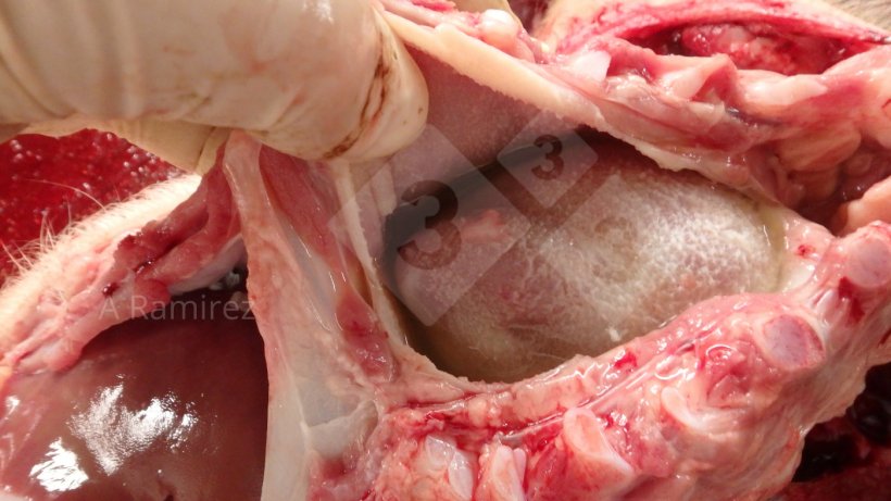
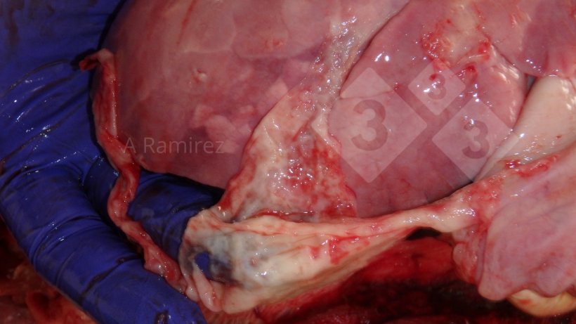
Scenario
Weaned pigs with polyserositis:
- For each pig affected, collect individual swabs of fibrinous exudate and place in Amies transport media and ship samples refrigerated to laboratory for bacterial culture, antimicrobial susceptibility, and PCR testing. Do individual testing on each sample; do not pool.


