

Always remember the vesicles, pustules and ulcers of FMD (Fig. 1), and the other vesicular diseases- inform authorities and wait.
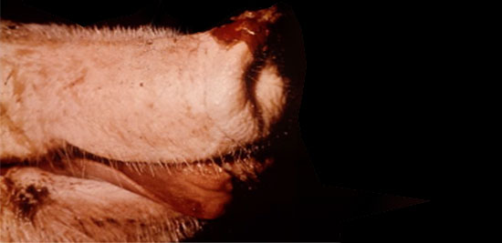
Figure 1 – FMD lesions on snout
Always remember that oedema under the jaw especially if accompanied by cyanosis and ulcerated lymph nodes and a line of inflammation may be Anthrax-smear and stain.
Swollen testicles and ulcerated scrotal skin may be Brucellosis or chlamydial infection.
In the first place, all the conformational abnormalities of the pig’s body are seen on or through the skin. So we may see wasting, fat accumulation, cysts abnormalities of muscles, joints, ligaments and tendons as well as whole body malformations such as Assymetric hindquarter syndrome, back muscle necrosis, pale watery muscles or pale dry muscles. Syndromes like leg weakness are all seen through the skin.
We have to rule out the congenital abnormalities and neonatal malformations eg joint ill or navel ill or abscesses or crushing by the sow. Neonatal one-off abnormalities may be seen (see Fig. 2) and more common conditions such as piglet anaemia may be seen in outdoor units where somebody has forgotten to open the pens to the earth (no supplemental soil or iron or iron injection).
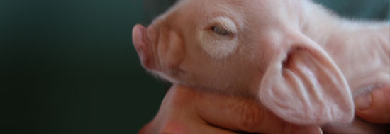
Figure 2 – Congenital abnormality “Dumbo pig with enlarged ears and periorbital oedema”
Dehydration, crushing and starvation (failure of maternal milk supply: too large a litter and bad simultaneous exposure of both rows of teats or blind teats or poor nutrition) of the neonatal piglet falls into these categories.
Epitheliogenesis imperfecta and Dermatosis vegetans, both rare, are specific skin abnormalities affecting the piglet. Thrombocytopenia and purpura are also often seen in this age of pig.
The second set of skin diseases refers to those manifestations that are caused by systemic disease. These often affect the systemic vessels and therefore cause changes in the blood-vascular system causing haemorrhages (petechiae, ecchymoses or frank haemorrhage), oedema in all the soft tissues especially the dependent parts, changes in blood flow such as congestion or hyperaemia resulting in colour changes in the skin.
The most common of these and often neglected is Erysipelas. Causes of sudden death should also be looked at as a differential (Anthrax, H. parasuis (see Fig. 3), electrocution, CSF, ASF, salmonellosis (see Fig. 4) etc). It can be seen as an acute case with the classical diamonds but more often than not they are not classical diamonds. Purple ears are often a feature. Bacteria can readily be isolated from the blood and lesions at post-mortem examination. The chronic cases may be more difficult to diagnose and may require P-M to look for joint and heart valve lesions.
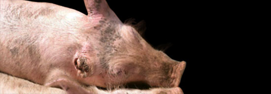
Figure 3 – Lost ear due to Haemophilus parasuis
| Figure 4 – Swollen red ears from Salmonellosis | Figure 5 – PRRS - “Blue ear” |
This second set would also include the deep blue/purple discolourations seen in anthrax, salmonellosis, PRRS (see Fig. 5) , P.multocida/ Actinobacillus pleuropneumoniae/H. parasuis septicaemias and particularly the two major haemorrhagic diatheses Classical and African swine fever. These are also confounded nowadays by Porcine Dermatitis and Nephropathy Syndrome (PDNS) which seems to occur as an acute dilemma as well as a more normal part of the PMWS syndrome. This is the biggest single worry for countries free from CSF or ASF that veterinarians do not recognise the potential occurrence of these diseases because they are concentrating on PDNS. This is precisely what happened in the UK in 2000 and has been an on-going possibility in other cases ever since.
Paleness (anaemia) is associated with either iron-deficiency in the newborn/neonate or M. haemosuis (Eperythrozoon) infection in the older pig.
Jaundice was rarely encountered in the pig until recently when it has become more common as a result of PCV2 infection causing hepatitis and hepatopathy.
Post-mortem examination will not help in the differential diagnosis of ASF, CSF and the haemorrhagic form of PDNS. The more you do, the more convinced you become of the practical difficulties of differentiation of the three conditions, LABORATORY DIAGNOSIS IS THE ONLY METHOD TO TELL THEM APART.
Often PDNS is the only option after the other tests and bacteriology for salmonellosis have proved negative. Only a proportion of PDNS cases will prove to have PCV2 virus as by the time PDNS occurs, often in excess of 16 weeks, the virus has disappeared


