

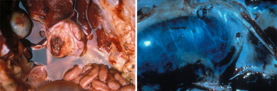
| Fig 1. ruptured bladder | Fig 2. kidney worms (Stephanurus dentatus) |
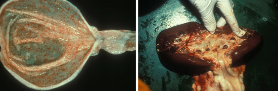
| Fig 3. cystitis | Fig 4. pyelonephritis |
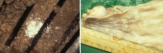
| Fig 5. crystals on the floor | Fig 6. uretero-vesical junction- normal |
FIG7 urethral orifice-crystalluria
The major diseases are as follows:
In the neonate we may find congenital abnormalities such as ureterocoele or ectopic ureters. However, the commonest can be kidney cysts and in many cases may be associated with cysts in the liver. There are also the possibilities of overlay or trauma from the sow.
In pigs of all ages we can find the serious diseases such as African Swine Fever, or Classical Swine Fever and other serious septicaemias such as salmonellosis or PDNS. There are over 30 different reasons for the classical turkey egg kidney: it is not pathogonomic for CSF or ASF. It is caused by congestion, or haemorrhage or emboli in the glomeruli.
White kidneys with streaks are often diagnostic for interstitial nephritis and this is often thought to be associated with leptospirosis.
Enlargement of the kidneys occurs in many disorders of the kidney. It is usually due to increased blood content and rarely urine and interstitial tissue. A reduction in kidney size is a result usually of chronic renal disease, and end-stage kidney. Alterations in shape may occur with cysts.
The colour of the kidney is altered by the degree of autolysis or hypostasis. These changes may be general(diffuse) or focal Increased redness may be associated with congestion associated with ASF or CSF, septicaemias or electrocution. Pallor may result from fibrosis , infarction, ochratoxicosis, anaemia, fatty change or tubular necrosis. Tan discolouration is seen in ochratoxicosis and brown colouration in porphyria. The focal reddening changes are seen in diffuse hyperaemia or emboli in the glomeruli. Focal pallor is a feature of infarcts or ischaemic necrosis or interstitial nephritis. White spots are usually indicative of the presence of interstitial nephritis. Not all white spots are leptospirosis but leptospirosis is the most commonly associated bacteria. Orange foci are found in the kidney when urate crystals or crystals are seen.
Twelve hours after death the kidneys start to soften and then become mushy(very soft). In chronic fibrosis they are tough, as they are in dehydration or neoplasia(nephroblastoma is not rare in young pigs). Gritty lesions may be seen in renal calculi. Gas bubbles can be seen in advanced autolysis. Haemorrhages that are severe are seen in warfarin poisoning or trauma. Ecchymoses are seen in ASF, or CSF, Salmonellosis, erysipelas, EMCV or Aujesky’s, slaughter, or poisoning, acute leptospirosis, pyelonephritis or endotoxaemia.
Alterations in the urinary bladder are generally restricted to the mucosa, and sometimes they extend into the submucosa. Nothing is pathogonomic except for the presence of inclusions in the mucosa. Mucinous degeneration may be seen occasionally. Haemorrhages in the bladder are seen in the major septicaemias. Mucus ,excessive debris, cells, bacteria, and fibrin are found in the lumen of the bladder as part of acute cystitis. In the sub-acute/chronic stages the neutrophils are replaced by lymphoid cells.
Microbiological investigation consists of culturing kidney tissue and urine but only fresh is any good. Should incubate on blood agar and MacConkey for 24 and 48hrs. Special horse blood plates are needed for Arcanobacterium (Eubacterium) suis. Acute cases can also be cultured for leptospirosis. E.coli is frequently cultured but no special features of these have been noted. Immunofluorescence is also used sometimes and negative ground illumination for leptospires. Porcine urine may show a variety of abnormal physical characteristics. These may include red urine, green urine, white or viscous urine, turbid, foaming, or ammoniacal smell. Various substances may appear such as pH changes, bilirubin, blood, glucose or ketones, and possibly nitrite or protein.
Sediments can be varied and include epithelial cells, erythrocytes, leucocytes, casts, bacteria, spermatozoa, crystals or mucus.
The three major disorders of the urinary system are Ochratoxicosis, Pyelonephritis associated with A. suis and Leptospirosis.
In ochratoxicosis the kidney lesions are very variable, with white streaks most commonly seen. There is also acute tubular necrosis. Testing for ochratoxin is possible but the half life is only 3-5 days. Levels in the feed of as low as 200 parts per billion will cause kidney lesions.
In cystitis and pyelonephritis there is often associated abnormalities of water supply, or locomotory problems. Diagnosis is usually post-mortem and requires isolation of the organism from kidney or bladder.
Leptospirosis as a herd problem is diagnosed by serology but individuals may require dark ground illumination, immunofluorescence, PCR techniques, or culture to prove infection.

