

Knowledge of the changing body composition of growing pigs is of vital interest to breeding companies, nutritionists and producers, in order to optimize production from different points of view (from nutrition adapted to the animal's needs to determining the optimum slaughter weight or obtaining the product desired by the pork industry or consumers). This composition can be determined in live animals with a computed tomography (CT) scan unit. CT is an emerging technology applied in animal production studies. CT is based on the emission of X-rays along the 360 degrees of the animal's body which, as they penetrate the body, are attenuated more or less depending on the density of the body tissues they go through. The implementation of reconstruction algorithms to the attenuation data matrices obtained allows us to obtain images from the inside of the animal. The advantage of CT is that it is a non-invasive technology, allowing us to determine the body composition of live animals (Figure 1) and, if necessary, at different times throughout the growth of the same animal.

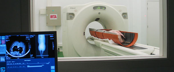
Figure 1: Examination of a live pig with a CT unit.
In IRTA, we have used CT scans to examine live pigs, of different genetics and gender, at different times of their growth (30, 70, 100 and 120 kg) through the project "In vivo evaluation of allometric growth of muscular and adipose pig tissues depending on the genetic and the sex by means of computer tomography" (INIA-RTA2010-00014-00-00). From the images obtained in specific anatomical areas, we have been able to determine different linear or curvilinear measurements and areas (fat thickness, loin area and perimeter, area and perimeter of fat, etc. — Figure 2). Also, by applying predictive equations based on images of the entire animal's body, the body composition (fat, lean tissue and bones) of the entire carcass was estimated, as well as that of its main cuts. This allowed us to study the evolution of different tissues depending on genetics and gender. For example, Figures 3 and 4 show, respectively, the allometric growth of fat and lean tissue within 4 different pork cuts —ham, loin, shoulder, belly and tenderloin— in pigs of different genetics and gender. The results show that, in general, the deposition of lean tissue (R2: 0.993 and root mean square error (RMSE): 0.486) was proportional to live body weight for all animals, regardless of genetics or gender. With regard to fat (R2: 0.994 and RMSE: 0.293), it is a late maturing tissue. In this case genetic differences were observed, with fat deposition in Landrace × Large White and in barrows happening later. Fat deposition in immunocastrated pigs is initially like that of boars, but after the second dose of the vaccine it is similar to that of females and barrows, therefore, the amount of fat deposited by the animal can be controlled by controlling the time of the second vaccination.
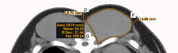
Figure 2: Linear, curvilinear and area measurements obtained on CT scans of the loin area (D = Top subcutaneous fat thickness, F = Lateral subcutaneous fat thickness, G = Loin area and perimeter.)
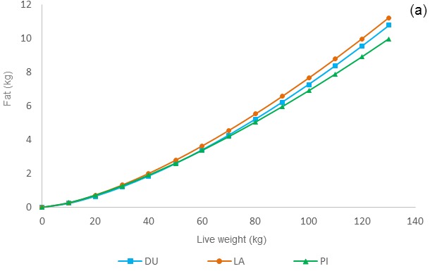
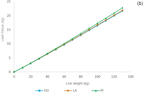
Figure 3: Allometric growth of fat tissue (a) and lean tissue (b) in the 4 main pork cuts —ham, loin, shoulder and belly— plus the tenderloin depending on the genetic cross (DU: Duroc × (Large White × Landrace); LA: Large White × Landrace; PI: Pietrain × (Large White × Landrace).)
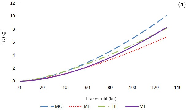
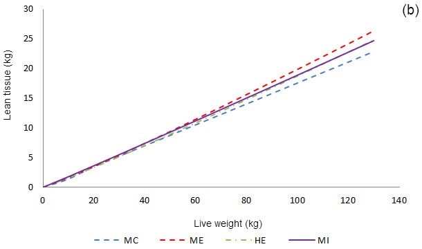
Figure 4: Allometric growth of fat tissue (a) and lean tissue (b) in the 4 main pork cuts —ham, loin, shoulder and belly— plus the tenderloin depending on the animal's gender (SCM: surgically castrated male; EM: Entire male; FE: female, IM: immunocastrated male.)
Work is currently being done on the evaluation of body tissues growth and bones characteristics depending on the pig's diet throughout project INIA-RTA2013-00040-00-00: "Influence of feeding restriction and dietary P levels on body tissue composition evaluated in vivo by computed tomography, bone mineralisation and sensory properties of the meat from gilts." In this project we'll use CT to study the effect of the feed restriction period (in volume or energy) on the deposition of fat and lean tissue, as well as the effect of the contribution of dietary phosphorus in bone strength.
Thus, the TC is a very useful tool in swine production as it allows us to model the growth of animal body tissues in vivo and to study the effect of different factors (genetics, gender, nutrition) on this growth.
