

Description of the farm
It is a nursery (site 2) that is part of a farm with a batch farrowing system in which 85-90 litters are weaned every three weeks (900-1,000 piglets/batch). The piglets arrive with an average age of 26-28 days and with an average weight of 7.4-7.6 kg.


Photo 1. Farm (outer view).
This nursery works in continuous flow (receiving a new group of piglets every three weeks), with at least two batches always present. The structure consists, basically, of three adjacent nursery rooms with all-in-all-out management (AIAO) per room. The pens, with around 29 piglets each, have concrete/plastic slatted floor, ad libitum feeding with linear troughs, and a suitable number of nipple drinkers. The ventilation is semi-natural with extractors and warm air heating. The hygiene and the managing standards can be considered as medium-high.
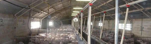
Photo 2. Nursery (interior view).
The health status is high (PRRS-, Aujeszky's disease-, App-, and atrophic rhinitis-negative). The piglets have been vaccinated against M. hyo in the farrowing rooms and, when arriving to the nursery they normally do not receive any treatment. In the past, some sporadic meningitis or sudden mortality problems due to Streptococcus suis have happened which have been successfully solved with amoxicillin in the drinking water (sometimes in the feed).
Until November 2011, the technical results in this nursery have been modest, with losses (dead and euthanized piglets) lower than 4%, an ADG higher than 450g, and a FCR of 1.6-1.7.
Appearance of the symptomatology, laboratory tests and diagnosis
.
Suddenly, by late November 2011, the owner detected an increase in mortality, with the loss of the "biggest piglets" in the older group and, according to the farmer, no particular symptomatology was seen, but every day some dead piglets were found.
In the first visit, the animals were active, with a good appearance, and they fed regularly. No obvious respiratory or digestive problems were seen. The necropsy, carried out in the farm on two animals, did not allow to draw any conclusion due to the absence of pathognomonic or indicative lesions. Samples of these two animals (meningeal swabs and intestines, spleen, and lung) were taken and sent to the laboratory, where the presence of Streptococcus or other bacteria that could cause “sudden death” as, for instance, haemolytic E. coli K88 (F4) was ruled out. By means of PCR and serological analyses it was confirmed that the site was still PRRS- and Aujeszky's disease-negative. As a preventative measure a treatment with amoxicillin (20 mg/kg LW) in the drinking water was prescribed, with no result. In the following days and weeks, the daily losses, apart from being recorded according to the batch, were recorded according to the pen, and it was seen that in certain pens, the mortality had reached 50%!.
Some days later, during the necropsy of other pigs, lesions (necroses) in the myocardium that could be indicative of Encephalomyocardovirus (EMCV) were shown.
Due to this, other samples (dead piglets and/or organs) were sent to the Lombardy and Emilia-Romagna Zooprophylactic Intitute's (IZSLER) headquarters in Brescia, and they confirmed the presence of the EMCV by PCR and cell culture. The Pathology Department of the University of Parma performed histological examinations of the brains and hearts of another three dead piglets. During the days immediately after, samples of faeces and urine were taken from piglets from several pens to verify if the animals could be virus excretors.
|
|
|
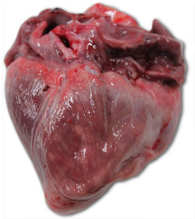
| Photo 3. Heart of a piglet that died at 40 days of age. We can see a great enlargement of the right ventricle, with multifocal necrosis (white patches) and gelatinous atrophy of the epicardial adipose tissue. | Photo 4. Piglet brain with congestion of the right cerebral hemisphere veins. |

|
Photo 5. Piglet brain. Thickening and congestion of the right cerebral hemisphere veins. |
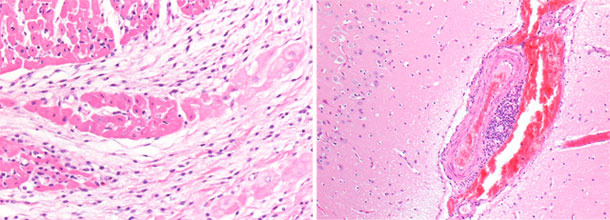
Photo 6. Myocardium, right ventricle. Basophilic focal necrosis of the myocardium with microvacuolization. |
Summary of the results
In spite of all the efforts to control the spreading of the infection, the losses (especially the sudden death of the biggest piglets) in group A went on, reaching 25%. This group, when taken to the fattening units still had a 7% of losses during the first two weeks in the new site. The losses in group B were reaching similar levels (18%) when, on December 1st, group C arrived to the nursery. The time sequence of the groups at the nursery are shown in the following diagram.

The pathology laboratory of the Veterinary Science Faculty at the University of Parma confirmed the diffuse presence of petechiae associated with discoloration patches in the myocardium, that are indicative of a serious form of myocarditis, and a diffuse congestion of the meninges.
The ten faeces and the three urine samples from groups A and B that were sent to the IZSLER, in Brescia, where all EMCV-positive.
Discussion
The virology and histopathological examinations confirmed that this serious case of sudden deaths, seen especially in groups A and B, was due to the infection with Encephalomyocardiovirus. According to the scientific literature, rats and mice are carriers and spread this infection. Although there are no data with respect to this, it is probable that a single family of rats/mice could trigger this problem initially. In fact, in this farm, the rodent extermination was very thorough and there were no evidences of a massive presence of rodents. No presence of rodent excrements was seen, and only after a detailed search some small mice footprints were found. So, there must be a period (surely brief) during which the pigs act as spreaders/multipliers of the infection. This period of time is probably of 1-3 days: the period that goes from T0 (moment of the infection) to the death of the animal. This hypothesis was confirmed due to the detection of EMCV in the faeces and urine and to the concentration of the mortality in certain specific pens.
Conclusion
Due to the high environmental contamination and to the high mortality (group B had reached 18% a bit more than a month after its arrival) in mid-December it was decided to empty the farm completely, taking all the piglets of groups B and C to an external fattening unit (site 3). Once emptied, the nursery was cleaned thoroughly using foaming agents, it was then cleaned with high-pressure hot water cleaners and it was disinfected. Although the control was already good, all the rodent extermination actions were checked and reinforced and, as an additional safety measure, all the rooms underwent a thermonebulization. The site was kept completely empty for a week. On December 29th a new group arrived and neither this one nor the following groups have shown a symptomatology compatible with EMCV.
Epidemiological notes
The occurrence of encephalomyocarditis virus (EMCV) among domestic pigs and wild boar in several European countries has been described and discussed in several articles. In a review done by Maurice H., et al (2005), found most clinical EMCV outbreaks were reported in Belgium (320), followed by Italy (110), Greece (15) and Cyprus (6). The outbreaks appeared to be clustered in 'endemic areas' with an increase in outbreaks during the autumn and winter months. The within-herd seroprevalence measured in clinically affected pig farms varied considerably among farms (2-87%), with age (0-84%) and by country. Data from farms with no clinical disease showed that subclinical infection with EMCV was found both within (seroprevalence 6-62%) and outside (up to 17 %) the endemic areas of the clinically affected countries as well as in the non-clinically affected countries Austria and France (3-5.4%). Among wild boar, the seroprevalence varied between 0.6 and 10.8%, and a study in Belgium found a prevalence of virus infection of 3.3%.


