Introduction
In June 2009 we are called for clinical problems on gilts in a newly established herd in Brittany. At the time of the call, 3 weeks before the arrival of the 3rd batch, some clinical signs appeared on gilts from the two first deliveries introduced in the farm:

- fever (temperature above 40.5°C)
- anorexia
- a relatively low fertility rate (86% for each of the first 2 batches of gilts inseminated, which was below the producer’s expectations)

Farm description and context
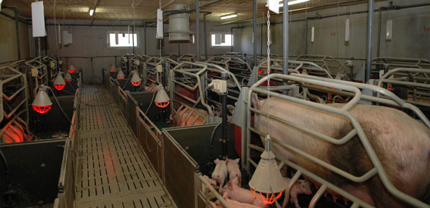
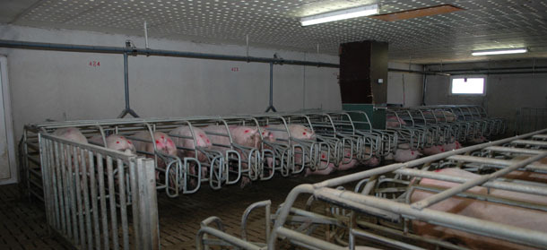
The 560-female farrow-to-finish farm is located in a high pig density area, in Brittany, and has begun herd population in January 2009. Gilts were introduced in 4 successive deliveries of F1 and / or GP (5th January, 9th February, 23rd March and 3rd May of 2009). The day of each delivery, blood samples were taken from 20 to 30 gilts in order to freeze the sera for possible analysis in the future.
The herd was managed by 10 batches, and piglets were weaned at 21 days of age.
Commercial semen was bought.
The farmer is extremely rigorous concerning both external and internal biosecurity measures.
The gilts F1 and GP came from 2 different nucleus farms of high health status. The farmer’s objective is to produce his own gilts.
The health status of the 2 nucleus farms is described below:
- PRRSv: negative
- Actinobacillus pleuropneumoniae: negative for all serotypes
- Aujeszky’s disease: negative
- Mycoplasma hyopneumoniae: negative
- SIV (H1N1, H1N2 and H3N2): negative
- PCV2: unknown status
The vaccination scheme on gilts at delivery included Parvovirus, Erysipelas and Swine influenza (H1N1 and H3N2).
A mange eradication program with ivermectin was implemented on gilts in quarantine.
Analysis of the situation and preliminary investigations
In June 2009, 3 to 4 weeks after clinical signs (fever and anorexia), 10 sick gilts were blood sampled.
They were found negative for PRRSv, SIV (IHA), Mycoplasma hyopneumoniae and leptospirosis (MAT).
Results of the 1st farrowings were bad:
| Alive born/sow | % dead born | % mummies | Weaned/sow | |
| Batch 1 | 11.67 | 2.8 % | 2.5 % | 9.94 |
| Batch 2 | 9.94 | 3.3 % | 7.9 % | 9.24 |
| Batch 3 | 10.23 | 2.7 % | 13.0 % | 9.51 |
Additional serological investigations on 25 sows confirmed the negative status regarding parvovirus, leptospiras and PRRSv circulation.
11 mummified piglets from 3 different litters were sent at the lab. The lab pooled the mummies per litter and samples were tested PCR for:
- PRRSv: all the results were negative
- PPV: all the results were negative
- PCV2: PCR results were found positive on hearts and livers with high titers varying from 4.2*109 to 8*1011 copies/g in the hearts and 3.6*1010 to 1.8*1013 copies/g in the livers.
Histological examination revealed small lesions of infiltration with mononucleate cells in the hearts but PCV2 antigens were seen in heart lesions by IHC.
► These results strongly suggested the role of PCV2 in the clinical signs observed.
PCV2 investigation by serology
It was thus decided to investigate the serological status of the gilts at arrival in the quarantine and if they seroconverted between arrival and the occurrence of the clinical signs.
Blood samples were collected at 2 time points:
- The day of arrival in quarantine (BS1): from 110 randomly selected gilts from the 4 deliveries
- After the outbreak of the clinical signs (BS2): 45 of the gilts were sampled a second time; these gilts were randomly selected regarding their physiological stage after outbreak of the clinical signs.
The presence of PCV2 antibodies was evaluated.
Mean PCV2 antibody titers were compared according to the batch and farm of origin. Homogeneity of titers was tested using a Student’s T-Test and a Kruskal-Wallis test. The statistical decision was made using a significance value of 0.05 (p<0.05).
Data at each time point (arrival in quarantine and after the outbreak of the clinical signs) were available for 45 gilts. Therefore, seroconversion was assessed only for these animals. PCV2 titers values and difference between 1st and 2nd sampling are presented in table 1.
Table 1. PCV2 antibody titre evolution (log10) and standard deviation (σ) at arrival in quarantine (BS1) and after outbreak of the clinical signs (BS2)
| Arrival in quarantine (BS1) | After the clinical outbreak (BS2) | Delta between BS1 and BS2 | |||||||
| n | mean ± σ | median | n | mean ± σ | median | mean ± σ | median | ||
| Delivery 1 | 22 | 0.19 ±0.318 | 0.000 | 22 | 3.10 ±0.543 | 3.04 | 3.10 ±0.543 | 3.04 | |
| Delivery 3 | 23 | 3.16 ±0.49 | 3.31 | 23 | 3.33 ±0.56 | 3.54 | 1.90 ±1.64 | 2.19 | |
All gilts of the 1st delivery seroconverted. In the 3rd delivery: 61.6% of F1 gilts and 60 % of GP gilts seroconverted.
► We can conclude that naïve gilts were introduced in the farm and that they developed clinical signs after having been in contact with PCV2 between their arrival in quarantine and the end of gestation period.
We were also interested in knowing the PCV2 antibody status at arrival in quarantine (details of the results according to the batch and farm origin are presented in table 2).
Gilts from the 1st delivery are negative for PCV2 antibodies at arrival in quarantine. All these gilts came from the nucleus farm producing F1 gilts.
Interestingly, some gilts from the same farm origin but introduced later (deliveries 2, 3 and 4) were positive (100% had titers above 1.3 log10 and 38.8 % had titers above 3 log10).
Furthermore, PCV2 serological status was different between the 3 deliveries: mean value of PCV2 antibodies of batch 3 was significantly above this one of deliveries 2 and 4 for F1 gilts (p=0.001), and the mean value was higher for GP gilts (p=0.107).
In all 3 deliveries 2, 3 and 4 there was no difference of PCV2 serological status between origins F1 or GP.
Table 2. Mean PCV2 titres (log10) at arrival in quarantine according to the delivery and origin in F1 as well as GP
| Origin | F1 (BS1) | GP (BS1) | Total | ||||||
| n | mean ± σ | median | n | mean ± σ | median | n | mean ± value | median | |
| Delivery 1 | 30 | 0.24 ±0.36 | 0.00 | 0 | - | - | 30 | 0.24 ±0.36 | 0.00 |
| Delivery 2 | 20 | 2.54 ±0.49 | 2.56 | 10 | 2.69 ±0.29 | 2.64 | 30 | 2.69 ±0.44 | 2.57 |
| Delivery 3 | 19 | 3.21 ±0.45 | 3.26 | 10 | 3.09 ±0.49 | 3.13 | 29 | 3.17 ±0.46 | 3.26 |
| Delivery 4 | 10 | 2.46 ±0.77 | 2.61 | 5 | 2.97 ±0.45 | 2.72 | 15 | 2.62 ±0.708 | 2.64 |
| Total | 79 | 1.82 ±1.36 | 2.44 | 25 | 2.91 ±0.44 | 2.72 | 104 | 2.83 ±0.574 | 2.72 |
Conclusion
First, we observe that despite the ubiquity of the PCV2, some animals coming from high health status farm may be naïve at 6 months of age, with no PCV2 antibodies. Secondly, it underlines a possible high variability of PCV2 serological status of gilts at the entry in a commercial farm: one delivery was negative, while the 3 successive ones introduced later were positive with heterogeneous titres values. At last, PCV2 serology results of one batch of gilts are not predictive of the subsequent batches.
Farmer’s objectives and actions proposed
In July 2009, a PCV2 vaccination program was implemented: the farmer was advised to mass-vaccinate the herd twice, 4 weeks apart.
Batches “at risk” (close to AI and close to farrowing) were vaccinated 2-4 weeks later.
Then, every booster was done 3-4 weeks before farrowing.
In September 2009, the rate of mummies dropped to an acceptable level (0.51 / sow). The number of still-born was correct.
The fertility rate was good.
| Mar 09 to May 09 | Sept 09 to Dec 09 | |
| Total born / sow | 11.83 | 12.46 |
| Mummies / sow | 1.34 | 0.23 |
| Still born / sow | 0.32 | 0.35 |
| Weaned piglets / sow | 9.42 | 11.4 |
| Fertility rate | 86% | 88% |
Discussion
The impact of PCV2 on reproductive disorders has been clearly demonstrated in experimental and field conditions. Most of field cases are related to naïve gilts.
This clinical case confirms the fact that PCV2 can be responsible for clinical signs and reproductive disorders observed in some gilts coming from farms of high health status.
The most interesting finding is the high individual variability of serological status of gilts entering a herd.
The diagnosis of reproductive disorders is still challenging and expensive but a complete differential diagnosis has to be performed. The impact of PCV2 on the performances of gilts and primiparous sows shouldn’t be underestimated. In the present farm, we never saw clinical sign of PMWS in piglets even in the piglets born during the reproductive disorders.
Mass vaccination of the herd twice 4 weeks apart, followed by boosters every 3-4 weeks before farrowing seems to be a good tool of prevention of clinical outbreaks. This vaccination is still maintained today and the health status of the farm is still high.