Farm description
The farm is a 1700-sow herd with external replacements. Gilts are acclimatised on the farm 60 days before their mating. Gilts are vaccinated against PRRS twice with a MLV, and they are PRRS ELISA positive and PCR negative. Replacement rate is 50%.

The farm is considered PRRS positive stable. The sow vaccination protocol includes a blanket vaccination every four months with a MLV PRRS vaccine and an Aujeszky's vaccine. Breeding animals are vaccinated against E. coli, parvovirus and erysipelas at every breeding cycle.
It is an old farm with successive refurbishments to allow better control and management of animals. The sows receive meal feed and the semen is bought externally.
Piglets are vaccinated against Circovirus and Mycoplasma at 21 days of age. Average weaning age is 23 days and average weaning weight 6,5 kg.
Case onset
Some weaners at the nursery started to show dyspnoea and persistent coughing. The number of “poor doers” rose close to an alarming 6%, with mortality above 4%. There is no response to in-feed medication (300 ppm amoxicillin and 80 ppm florfenicol).
The respiratory clinical signs worsens when the animals are moved to the grower/finisher barn (4% mortality). Some animals showed cachexia and ear tip necrosis. Some of them even show the "dog sitting" position, and between 16 to 20% have strong dyspnoea and anorexia.
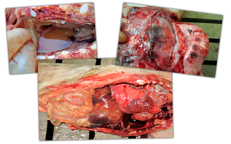
Figure 1: Thoracic cavity - Lesions in a nursery pig: pericardial oedema, multifocal pulmonary lesions and septicaemia with respiratory and digestive picture
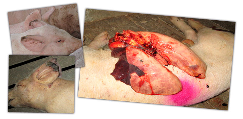
Figure 2: Lesions in a growing pig: ear tip necrosis and pneumonia in the cranial and medial lobes.
Lesions and clinical signs must be taken into account in order to obtain a differential diagnosis
As well as the identification of potential pathological agents, a crucial step to determine the farm’s clinical problem was to perform a breakdown of the different environmental and management agents, and their impact on respiratory pathologies.
The lesions found at necropsy made us suspect that the farm is facing a multifactorial porcine respiratory disease complex (PRDC).
Porcine respiratory disease complex (PRDC)
Porcine respiratory disease complex (PRDC) is a multifactorial disease where interaction of host –infectious agent is combined with environmental characteristics.
The infectious agents involved in PRDC can be classified as:
Primary agents : PRRS, Influenza, PCV-2, Aujeszky and Mycoplasma hyopneumoniae
Secondary agents: Streptococcus suis, Actinobacillus suis, Actinobacillus pleuropneumoniae, Pasteurella multocida y Bordetella bronchiseptica. Mycoplasma hyopneumoniae can also act as a secondary agent.
Environmental and management elements that may have an effect on the respiratory complex are listed in Table 1.
Table 1. Role of environmental and management factors on respiratory pathologies.
| Factor | Optimum value | Effect |
| Temperature | 25-40 kg LW ⇒ 18-22 ºC 40-100 kg LW ⇒ 15-22 ºC |
Excessive and insufficient values affect ADG, FCR, and predispose to the appearance of pathologic conditions. |
| Ventilation | <0,2 m/s | Excessive ventilation could predispose to pathologic conditions. |
| Pig Density | 20-30 kg ⇒ 0,3 m2 30-50 ⇒ 0,4 m2 50-85 ⇒ 0,55 m2 85-110 ⇒ 0,65 m2 |
Minimum values set by law. Better conditions give better performance. |
| Group size | <20 animals/pen | |
| Feed troughs | Good regulation at the entrance (not feed excess). Empty them at least twice a week to avoid accumulation of feed fines. | |
| Barn orientation | East-west | Achieving best environmental conditions in summer and winter. |
| Insulation | Proper insulation improves performance and helps to prevent disease. | |
| Slurry Pit | In some situations it is useful to add some water to avoid draughts. | |
| Water pipes | Cleaned with organic acids to avoid growth of "biofilm" together with the medication system. |
|
| Environmental pollutants | NH3 ⇒ <20 ppm CO2 ⇒ <3000 ppm CO ⇒ <10 ppm H2S ⇒ <0,5 ppm |
The combined action of these agents and dust, increase susceptibility to respiratory problems. |
Investigation protocol
After evaluating clinical signs and lesions, a comprehensive approach was adopted to find out which agents played a more important role in the development of the disease.
To determine the agents involved, the following laboratory tests were performed:
Serology
- Piglets at birth before colostrum intake to evaluate PRRS circulation in gestation. 10 samples
- > 21 day-old “poor doers”, to evaluate PRRS recirculation in the farrowing house. 10 samples
- Weaners - 6 and 9 weeks of age to study the dynamics of the disease at the nursery. 10 sera per age group
Microbiology, culture and sensitivity, and histology:
- Submission of lungs to the laboratory: 10 - 12 lungs every 2 or 3 weeks.
Results
PRRS Serology
In the serological tests, piglets at birth (before colostrum) were PCR negative, but samples from 21-day-old runts at the farrowing house were 100% PCR positive. PCR was also positive for 6 and 9-week-old animals. Those results indicate that there is an infection in the farrowing house and the virus re-circulates at the nursery. However, the farms does not show any reproductive clinical sign.
Microbiology
Only one opportunistic pathogen (Aerococcus viridans), which did not fit the clinical picture, was isolated from the lung tissue culture. The reason could be that samples came from medicated animals since, when a clinical problem arises, animals are always in-feed medicated until the antimicrobial results arrive.
Treatment
After reviewing farm routines, it was concluded that All In-All Out flow (AI-AO) was not respected, with runts and low weight animals kept for more than 35 days at the farrowing house because they did not reach the 6,5 kg selling weight. To try and correct this problem, piglets were identified with a different tag colour depending on their birth week, which make control much easier, therefore achieving a better implementation of strict AI-AO flow.
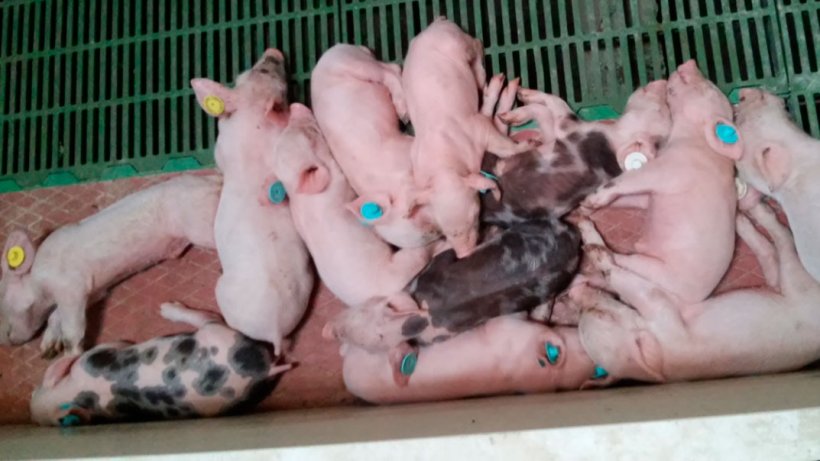
Figure 3: Different age runts in the farrowing pen. Piglets tagged according to birth week.
In addition to reviewing piglet management at the farrowing house, blanket vaccination with a MLV PRRS vaccine was performed at the farm. Biosecurity protocols were reviewed increasing control on transport, visitors, veterinarians, maintenance staff etc
However, after all this protocol was implemented, problems at the nursery and grower/finisher section remained. As the response to antibiotics was still very poor, some other viral process associated with PRRS became major suspects.
There were no problems, and there had never been before, at the sow farm. Measures taken so far (PRRS vaccination, improved biosecurity) were not sufficient to solve the problem. Therefore, a second study of risk and predisposing factor was performed.
Second investigation protocol
- Cleaning procedures were reinforced. Washing and disinfection protocols were changed.
- Strict AI-AO flow was implemented and monitored at the nursery trying to displace PRRS virus.
- The use of local and full space heating systems in the grower facilities was implemented. The goal was to avoid temperature fluctuation and temperatures under 24 degrees.
After implementation of these measures, the flow of animals at the farrowing house and nursery became PRRS negative by PCR. However, there were still a 4% of "poor doers" at the nursery (from the initial 6%).
Respiratory problems continued at onset of growing period, but response to antibiotics was good. Even so, there was still a great weight variation. The strong temperature variation could be a stress factor, triggering a multifactorial process. The plan was to take further samples to check that there were no more pathogens, such as influenza, taking the role of primary pathogen and causing secondary bacterial infections that could lead to the chronic respiratory disease seen at the farm.
Table 2: Influenza PCR test - Oral fluids .
| Haemagglutinin | Neuraminidase | |||
| Sample | H1 | H3 | N1 | N2 |
| y/n | + | - | + | - |
Samples were positive to H1N1 serotype.
Treatment
The whole breeding herd was vaccinated twice against Influenza and this was included in the sow vaccination schedule for every cycle. Strict implementation of AI-AO flow of piglets was achieved at the sow farm and the nursery. The aim was that the passive immunity transferred from sows to piglets would significantly reduce virus re-circulation and, therefore, transmission ratio, thus achieving a negative flow.
Continuous flow involving significant age differences is always very challenging in terms of respiratory pathologies.
At a later stage, when piglets from vaccinated sows arrived, together with an adequate flow of animals, the problem was eventually corrected in nursery and finisher pigs. Medication expense was significantly reduced in site II and III, and mortality levels were reduced to less than 2.5% in nursery piglets and 2% in finishers. By controlling Influenza, secondary pathogens became less able to cause severe lung injury.
Therefore, when approaching a disease process, the whole pyramid must be monitored until the agent playing a primary role at the onset and development of the infectious process is identified. We learnt that poor environmental conditions, together with the pathogen, increase the severity of the problem.
Conclusions
- An integral approach needs to be performed in order to reach a correct differential diagnosis, confirming which pathogens play an important role in the development of the clinical signs seen at the farm.
- Laboratory tests are a tool, not the diagnosis.
- Proper implementation of AI- AO systems avoids exposure between animals of different ages (commingling different age groups is a big problem). It is necessary to establish management protocols that limit continuous flow and, as much as possible, large AI-AO production flows. It is the only way to limit infectious processes.
- Improvement of environmental factors (reducing stress improves the animal’s immune system): optimize the animals well-being by means of providing good standards on room temperature, ventilation and water.
- Most pig clinical diseases are multifactorial and the clinical resolution goes through the modification of different aspects.