

All of us should consider that some of the major epizootic pig diseases have as a component nervous diseases as a result of systemic involvement- eg Aujesky’s disease (neurons), Classical Swine Fever or African Swine Fever (blood vessels of the CNS). Most of these can be detected by clinical signs, notably by use of the thermometer and the demonstration of high pyrexia, but also by the presence of signs in other systems and obviously by attention to laboratory techniques.
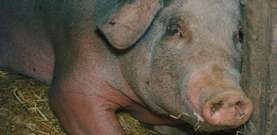
A pig with CSF
There are three forms of Aujesky’s disease –respiratory, reproductive and neurological and this last is the most serious and dramatic.
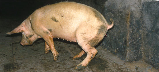
Aujesky’s: lame pig
Teschen has disappeared from the list of the major epizootics, quite rightly as it only appeared as a serious disease in that one part of Czechoslovakia which gave its name to the disease (seen as pyrexia, stiffness, tremors, convulsions, paralysis and death in 3-4 days). Milder strains were called Talfan. These can still occur and together with all the other minor enteroviruses of pigs are all best regarded as porcine enteroviruses. These enteroviruses have wide variation in strains and therefore pathogenicity. These can cause quite serious encephalitis and myelitis in susceptible pig populations and in these cases the virus needs to be isolated from the spinal cord or brain lesions. Histology needs to be carried out on brain and spinal cord and if necessary fresh tissue kept for micro-array analysis. There may be quite serious perivascular cuffing with neuronal necrosis, malacic foci, chromatolysis and neuronophagia. The lesions are usually more severe in the grey matter than the white matter, and the motor neurons of the ventral horns of the cord are more affected than those of the dorsal horn.
Having ruled out the serious epizootics what are we left with. I am a great believer in the old saying that common things are common and this is where we should start. The most likely clinical diagnosis in young pigs is Streptococcus suis infections usually type 2 or 1 or 1/2 but rarely other serotypes.
If there are suddenly groups of young pigs dying or showing nervous signs but which respond well to penicillin, if there are pigs with swollen joints, or pigs with pleurisy, pericarditis or peritonitis then a post mortem examination and differential laboratory diagnosis by culture and serotyping will tell you if it is S. suis or possibly H. parasuis. In some cases the the presence of S.suis is suggested by the herniation of the cerebellum into the foramen magnum caused by the brain swelling. A swab inserted through the foramen magnum will enable you to recover S. suis from the meninges which may well be opaque in the classically affected case.
Similarly mild infections with low morbidity and mortality in pre-weaned pigs with vomiting, wasting, and lethargy but quite often good recovery may be associated with Vomiting and Wasting Disease Virus, more properly called Haemagglutinating Encephalitis virus. This is widely present in pig populations and usually because of high exposure does not cause a problem.
In some areas there may be a problem in young and post-weaned pigs of Encephalomyocarditis virus (EMCV) which is a rodent virus that causes both neurological and cardiological signs in pigs. Post mortem examination, and the gross appearance of white streaks in the heart indicating possible necrosis, mononuclear cell infiltration and fibrosis together with oedema in the body cavities is strongly suggestive of EMCV. Collection of histological samples from the heart muscle will enable a confirmatory diagnosis and confirm the presence or absence of mulberry heart disease at the same time which may present with similar cardiothoracic lesions. The virus can be confirmed by PCR, RT-PCR or by culture of the virus.
Nowadays, the likelihood of the old style of poisoning that could occur with the accidental overdosing of drugs or mixing in of dangerous compounds is unlikely. Thus incidents of arsanilic acid poisoning, organophosphorus poisoning and chlorinated hydrocarbons are not reported. However, they may still occur as may plant poisonings such as bracken, but the one likely to possibly still occur in a home-mixing situation is with selenium as the magnitude of change between a therapeutic level and a toxic level are quite small. A large outbreak affecting over 6000 animals in the UK in the 1980’s was believed to be due entirely to the mis-reading of the place of the decimal point so 20kg was added instead of 2kg. Care has to be taken when mixing any rations and most of all if you are home –mixing.
There are two other quite common conditions that are found as a cause of neurological conditions in pigs which are in fact not related to neuropathic agents but which are the result of toxic changes in the brain. The first of these occurs commonly 5-7 days after weaning and is associated with proliferation of certain strains of E. coli in the gut which produce enterotoxins that are absorbed and damage the brain causing amongst other changes angiopathy of the small vessels. A bilateral line of malacia may be seen in the medulla through to the mid-brain. Diagnosis is by timing of clinical signs, oedema of mesentery, greater curvature of the stomach or eyelids and isolation of the pathogenic O serotypes from the gut.
 |
 |
|
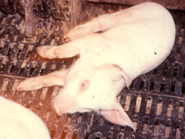
Oedema disease affected piglet 10 days after weaning
|
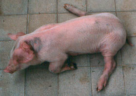
Oedema disease: affected pig
|

