

Accurate diagnosis of disease when pigs are showing clinical signs suspicious of PRRSV is important to allow suitable control measures for both PRRSV and other diseases occurring at the same time. The methods used are discussed below.
Clinical signs:
may mean that PRRSV is strongly suspected, further investigation is essential for a definite diagnosis to be made.Non breeding pigs:
• wide variety and nonspecific signs; in laboratory submissions with PRRSV from 2003-2009 to theReproductive disease:
Veterinary Laboratories Agency (VLA) in the UK, the four most common signs were respiratory, wasting, malaise and found dead.
• signs not readily distinguished from other common diseases of rearing pigs. PRRSV contributes to
the porcine respiratory disease complex (PRDC) and also increases the severity of other diseases on farm such as streptococcal meningitis.
• PRRSV often suspected where there is an upsurge in respiratory or other disease of unusual
severity, or which is not responding to antibiotic treatment as well as expected.
• characterised by late abortions, stillbirths, weak neonates, variable piglet and litter sizes, increased preweaning mortality and irregular returns.
• litters may show evidence of sequential foetal death with mummies, stillbirths, weak live and viable piglets born to the same sow.
• sows and gilts may show few signs or transient inappetance, malaise and pyrexia, and occasionally deaths.
• coughing may occur, particularly in younger breeding pigs, such as replacement gilts.
• in association with the poor viability of piglets at birth and the effects of disease on sows’ milk supplies, preweaning mortality may increase with mixed and unusual causes of death.
• severity of disease very variable due to differences between virus strains, different levels of immunity in the pig herd (from vaccination or previous exposure) and herd specific factors affecting when pigs are infected and how the virus spreads within the herd.
• disease can be sufficiently severe to warrant reporting to the Animal Health Authorities if the signs are indistinguishable from notifiable diseases; classical swine fever and Aujezsky’s disease.
Clinical signs reported in growing pigs with PRRSV submitted to VLA 2003-2009
Post-mortem examination (PME) - pigs for PME should be typical and early cases of disease submitted to the laboratory live (if welfare allows), or freshly dead, and ideally untreated. Chronic cases from hospital pens are unlikely to be useful. PME alone does not allow PRRSV to be diagnosed but is a good starting point in investigation because:
• allows pathology (disease processes) in the pig’s different organ systems to be assessed and provides excellent material for testing to diagnose, or rule out, PRRS.In reproductive disease, submission of several whole affected litters is advisable to provide material for diagnosis of PRRSV and other diseases causing reproductive or neonatal problems. If available, fresh stillborn and weak neonates are more useful than decomposing aborted foetuses.
• PRRSV commonly present with other diseases, PME allows full investigation of these.
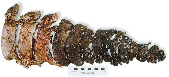
Progressive foetal death in a litter can be seen in PRRSV infection
Polymerase chain reaction (PCR):
a very useful sensitive method of directly looking for PRRSV in the tissues or blood of a pig. Most methods distinguish Genotype 1 (European) and Genotype 2 (North American) PRRSV.• A positive result indicates that the pig was actively infected with PRRSV at the time of sampling. Assuming that the pig was not recently vaccinated with live vaccine, this confirms that field PRRSV infection has occurred. On a unit which should be PRRSV-free this provides confirmation of PRRSV. However, as PRRSV is present in serum and tissues for an extended period (weeks), further investigation (IHC) is often undertaken to assess how much the PRRSV was contributing to disease, especially on units where PRRSV is known to be present.
• In foetal or neonatal tissue, a positive result is confirmation of PRRSV. A negative PCR result does not rule out PRRSV as the virus can infect the foetus and cause damage, but the virus may have gone by the time the piglet is delivered. It can be useful to perform PCR on serum from affected sows in reproductive disease, collecting blood from sows showing signs, not historical cases. This may also allow virus to be detected.
Immunohistochemistry (IHC):
histopathology and lung IHC determine whether PRRS virus is causing lung damage by labelling the virus within lung tissue. It can be used alongside other tests to investigate the contribution of PRRSV to pneumonia relative to bacteria (Pasteurella multocida, Actinobacillus pleuropneumoniae, Haemophilus parasuis, Streptococcus suis), Mycoplasma hyopneumoniae and other viruses (PCV2, swine influenza). This test is particularly useful in pigs which are vaccinated for PRRSV or pigs from herds where there is a known PRRSV challenge but infection has previously been controlled. It is essential that pigs are submitted early in disease and are not longstanding cases for this to be useful. Where pigs are PCR positive, and lung IHC negative, the presence of PRRSV is still of significance in sick pigs as it can be causing immunosuppression. It is not generally used for foetal tissues.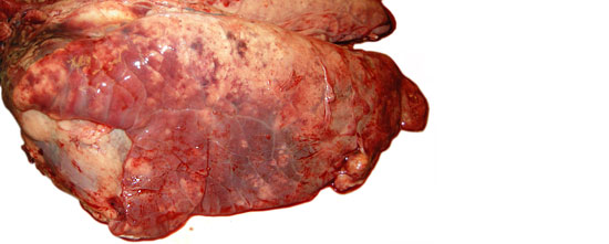
Pneumonia in 14 week old pig with PRRSV viraemia, PMWS and Pasteurella multocida infection; although PRRSV was not identified in lung by IHC it is likely to be exacerbating PCV2 disease in this pig.

