There are three primary groups of rotaviruses which cause diarrhea in pigs and they include Rotavirus A, Rotavirus B, and Rotavirus C. Each group of virus is different from each other and requires specific assays designed for them. Pig diarrhea often involves infections with multiple groups of rotaviruses at the same time.
Assays available
Gross pathology

- Evaluates the presence of small intestine wall thinning which can suggest the presence of disease.
- Location of lesions: jejunum and ileum
- Pros:
- Inexpensive and quick results
- Applies to all three groups of rotaviruses
- Cons:
- Not diagnostic – similar lesions can be found with other diseases especially coronavirus infections (transmissible gastroenteritis, porcine epidemic diarrhea, and porcine delta coronaviruses)
- Need fresh tissues (<15 min) as decomposition dramatically changes the appearance of small intestine
- Unable to differentiate between different groups of rotaviruses
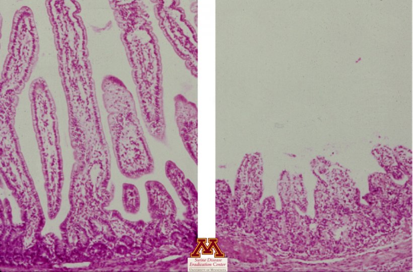
- Evaluates the presence of tissue lesions (small intestinal villus atrophy) which can strongly suggest the presence of disease
- Sample types: jejunum and ileum tissue
- Pros:
- Confirm villus atrophy suggesting viral enteritis
- Cons:
- Need to formalin-fix intestinal tissues within <15 min of pig death -Intestinal tissue decomposes quickly
Immunohistochemistry (IHC)
- Detects presence of viral antigen
- Sample types: jejunum and ileum tissue
- Pros:
- Definitive diagnosis: can confirm presence of virus with associated lesions
- Cons:
- Currently only available for Rotavirus A and Rotavirus C (not available for Rotavirus B)
Polymerase chain reaction (PCR)
- Detects presence of specific sequence of viral nucleic acid (RNA)
- Sample types: jejunum and ileum intestinal tissues, fecal swabs, feces
- Pros:
- Multiplex PCR can detect different rotavirus groups including Rotavirus A, Rotavirus B, and Rotavirus C
- High sensitivity
- PCR quantification
- Moderate cost
- Can often do pooling of feces or tissue samples to lower cost while minimizing loss of sensitivity (especially regarding clinical relevance).
- Cons:
- High sensitivity – can confirm presence of virus but not disease (high prevalence of virus in environment)
Virus isolation
- Isolates live virus
- Sample types: jejunum and ileum intestinal tissues, fecal swabs, feces
- Pros:
- Isolate virus for use in vaccine development (autogenous vaccines)
- Cons:
- Expensive
- Slow results
- Currently only available for Rotavirus A and not available for Rotavirus B or Rotavirus C
- Often difficult to grow (many false negatives)
- Sampling handling from field to laboratory can impact virus survival
Enzyme-linked immunosorbent assay (ELISA)
- Detects presence of antibodies
- Sample types: serum
- Pros:
- Animals remain positive for several weeks
- Can identify negative herds
- Cons:
- High prevalence of virus in environment results in common exposure to many/most pigs
- Need assay specific for each rotavirus group
- No cross protection between different groups of rotaviruses
Table 1. Summary of the applicable assays with respect to the different rotavirus groups.
| Rotavirus A | Rotavirus B | Rotavirus C | Can differentiate between groups? | |
|---|---|---|---|---|
| Gross pathology | Yes | Yes | Yes | No |
| Histopathology | Yes | Yes | Yes | No |
| Immunohistochemistry (IHC) | Yes | No | Yes | Yes |
| Polymerase chain reaction (PCR) | Yes | Yes | Yes | Yes |
| Virus isolation | Yes | No | No | No |
| Enzyme-linked immunosorbent assay (ELISA) | Yes | No | No | No |
Result interpretation
Gross pathology
- Positive: Preliminary confirmation of the presence of intestinal thinning
- Negative: Early or mild cases may not manifest with extensive intestinal lesions
Histopathology
- Positive: Confirmation of disease, but not cause of disease
- Negative: Negative, or lesions could have been missed if testing wrong sample or too late after infection
IHC
- Positive: Virus is present at site of lesion
- Negative: Negative, or virus could have been missed if testing occurs too late after infection or the concentration of virus is too low (only present for a very short period of time). Does not detect Rotavirus B.
PCR
- Positive: Confirmation of presence of the different groups of rotaviruses (A, B, and C)
- Negative: Negative, virus could have been missed if testing occurs too late after infection or poor sample selection or handling
Virus Isolation
- Positive: Confirmation of presence of virus but not disease
- Negative: Negative, virus could have been missed if testing occurs too late after infection or simply unable to grow due to other contamination or poor sample handling (present but can be difficult to culture). Does not detect Rotavirus B or Rotavirus C.
ELISA
- Positive: Past exposure (> 2-4 weeks) to vaccine or wildtype virus. No association of positivity and disease.
- Negative:
- Negative to vaccine or wildtype virus
- Infection too early to detect
Scenarios
Neonatal pigs with scours (acute or chronic)

- Collect fecal samples from 10 or more untreated scouring pigs and submit for PCR testing for Rotavirus A, Rotavirus B, and Rotavirus C in pools of 5.
- Necropsy of 1-3 recently dead or euthanize scouring pigs. Grossly evaluating jejunum and ileum for thinning of intestinal wall and collect samples and place in formalin solution for histopathology and IHC evaluation.
Early post weaning pigs with scours (acute or chronic)
- Collect fecal samples from 10 or more untreated scouring pigs and submit for PCR testing for Rotavirus A, Rotavirus B, and Rotavirus C in pools of 5.
- Necropsy of 1-3 recently dead or euthanize scouring pigs. Grossly evaluating jejunum and ileum for thinning of intestinal wall and collect samples and place in formalin solution for histopathology and IHC evaluation.


