

Although pigs can be infected with different species of coccidia, in suckling piglets the most important disease caused by enteric protozoa is coccidiosis due to Isospora suis.
Biological cycle
I. suis has a cycle that is important to know in order to understand the diagnosis, the treatment, and the prophylaxis of coccidiosis. In this cycle, I. suis has an ambient phase (oocyst) that, in adequate conditions, can remain viable for up to a year, which facilitates the infection remaining present in farrowing pens.
 |
|
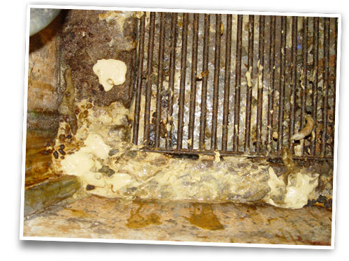
Pasty yellow feces
Author: Jesús Bollo
|
The infected piglets eliminate these oocysts by way of the feces in a non-sporulated form, therefore they are not infectious. In order to become infectious, the oocysts have to sporulate, and the time they take to do so depends on the temperature conditions and the humidity of the environment they are in.
In the farrowing pens, generally the conditions for sporulation are quite adequate. The I. suis oocysts sporulate quickly between 20°C and 37°C and the heat of the warming plates in the pens makes it so that the oocysts can sporulate within 12 hours.
The oocysts are more sensitive to inactivation before and during sporulation. Once sporulated, they are extraordinarily resistant to environmental conditions and disinfectants.
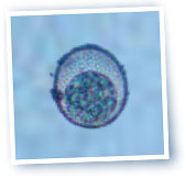
During sporogony, the I. suis oocyst forms sporocysts, each one of which with four sporozoites.
A receptive piglet is infected orally upon ingesting sporulated oocysts. Passing through the stomach, the digestive enzymes and bile salts activate the sporozoites. These sporozoites are set loose into the intestine and penetrate the enterocytes, begining the asexual multiplication phase, or schizogony, in the cytoplasm of these cells throughout the entire small intestine, but mainly in the jejunum and the ileum. Asexual multiplication mainly takes place in the enterocytes of the tips of the intestinal villi.
This asexual multiplication has two phases. In the first, the sporozoites that have invaded the enterocytes transform into schizonts or type 1 binuclear merozoites. Within 24 hours each nucleus turns out one type 1 infectious merozoite, which will penetrate the other cells to repeat the multiplication process. The second phase of asexual multiplication turns out type 2 merozoites.
These type 2 merozoites transform into macrogametes (female gametes) or microgametes (male gametes), that produce, in the infected cell, microgametocytes and macrogametocytes.
In the sexual reproduction phase, the macrogametocytes fertilized by microgametocytes are transformed into a zygote which forms a non-sporulated oocyst which is eliminated by way of the feces in order to restart the cycle.
Pathogenesis and clinical signs
The infected enterocytes are destroyed, leaving the lamina propria exposed, and are replaced by immature enterocytes that don’t have the same capacity to digest or absorb that mature enterocytes do, giving rise to malabsorption and fluid loss. The hyperplasia of the crypts also contributes here, with its secreting activity in diarrhea.
The incubation period is 3-4 days, depending on the infecting dose, and the clinical signs manifest more intensely in piglets 7-10 days old and appears throughout lactation but less often in weaned piglets.
The mortality rate is variable and, without treatment, can reach 20%. In farrowing pens, not all litters are affected, and the severity of the illness can also differ greatly between piglets from the same litter.

 |
|
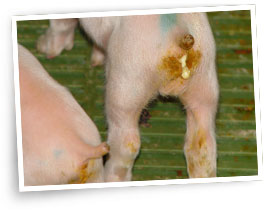
Fecal-stained perineum
Author: Jesús Bollo
|
The main clinical sign is a yellowish or grayish diarrhea that doesn’t respond to any antibiotic treatment. The feces are pasty at first and become more and more liquid as the illness progresses.
The piglets are wet and dirty with feces, they have an odor similar to that of rotten milk and their hair is long and bristly. The fact that these piglets remain wet leads to hypothermia and contributes to the severity of the disease. The disease causes dehydration and weight loss.
The presence of other concurrent infections can aggravate the clinical picture and increase the mortality rate.
Lesions
In the small intestine, specially in the jejunum and the ileum, there is an enteritis that varies between catarrhal in mild cases to fibrinonecrotic in piglets with severe clinical signs. The intestinal content is pasty or aqueous with necrotic flakes.
Microscopically there are atrophic lesions, fusion of the villi, hyperplasia of the crypts and necrotic enteritis.
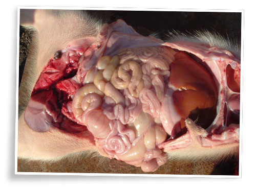
Author: Librado Carrasco
Diagnosis
Clinically, coccidiosis is suspected when there is diarrhea in lactating piglets, above all from 7 to 14 days of life, which does not respond to antibiotics.
In the laboratory this is confirmed by the detection of oocysts in feces, combined with the detection of different phases of the cycle of this parasite in mucous scrapings from the jejunum or the ileum.
Treatment and control
In coccidiosis caused by I. suis the importance of the sow feces as a source of infection for the piglets is limited (she is an asymptomatic carrier); the susceptible piglets are infected mainly by contaminated farrowing pens that haven’t been sufficiently cleaned and disinfected, which permits viable oocysts to remain in the environment.
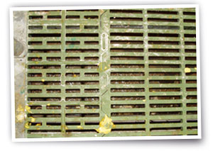
Farrowing pens should be thoroughly cleaned, all the organic material should be removed and they should be disinfected with sodium hypochlorite or some derivative thereof, although most often used are derivatives of ammonia.
Also, it is very important that the pen is very dry since oocysts are very sensitive to dryness. In serious cases, disinfecting and flame-drying the floor is recommended, if the materials of the pen will permit.
Author: Jesús Bollo
Given that the main sources of infection are contaminated farrowing pens and sick piglets, the preventive treatment of the pigs is not very efficient, although anticoccidials such as decoquinate and salinomycin can be used.
Currently, the most effective drug against I. suis is toltrazuril, which is active when faced with sexual and asexual phases of the cycle of the coccidian.
In piglets, the administration of toltrazuril is efficient as prophylaxis as well as treatment. The prophylactic application of one single 20 mg/kg p.v. dose at 3-4 days old obtains a considerable reduction of prevalence, clinical profile, and the elimination of oocysts in affected piglets.
The same dose is administered for the treatment of affected piglets, and is also highly efficient.