Farm introduction
The farm in this case study is a large breeder-multiplier farm located in the pig-dense areas of north-east China (Dongbei), near Shenyang, in Liaoning province. The farm system was established 3 years ago, in new construction buildings on a large single-site within a perimeter fence and boundary.

Pigs are weaned from all gilts and sows at 21 days old, and then raised in adjacent nursery buildings. At around 10 weeks-old, pigs are moved to separate grow-out finisher buildings. The farm has a large work-force. There is also a small team of technical and veterinary experts, with some outside consultants. The farm system purchased compound feed from a large feed mill operation. Finisher pigs are sold locally to live markets, dealers and slaughterhouses. Any dead pigs are collected by trucks for disposal by local firms.
The breeding sows and gilts received commercial vaccinations against E. coli, parvovirus, erysipelas and leptospirosis. Piglets are vaccinated at various points between 2 and 8 weeks-old against pseudorabies (Aujesky’s disease), Mycoplasma and FMD (foot and mouth disease) with commercial vaccines.
Like many farms in China, the nursery mortality rate generally occurred at 8 to 15 %, mainly due to PRRS and related secondary conditions. The levels of mortality in the finisher and breeder sheds were generally much lower (0 to 3 percent).

Emergence of the case
From May to June 2018, the breeder farm area had been re-stocked with several batches of incoming gilts up to a total of 4,500 adult age pigs (greater than 3 months-old). These batches of pigs had come in several truck deliveries from local north-east breeding company origins.
From May to August 14th 2018, 18 of the 4,500 pigs had been culled or died, due to various endemic conditions (less than 1 per week).
On August 15th, several pigs in one building were noted to be depressed with a high fever, bloody discharges, with 3 deaths.
On August 16, 10 sick pigs were noted, with 8 deaths
On August 17, 150 sick pigs were noted, with 23 deaths
On August 18, 300 sick pigs were noted, with 26 deaths
By August 19, a total of 615 sick pigs and 88 deaths were counted.
The case situation therefore emerged quickly, with large numbers of dead and dying pigs discovered by workers in the sheds over a 4-day period.
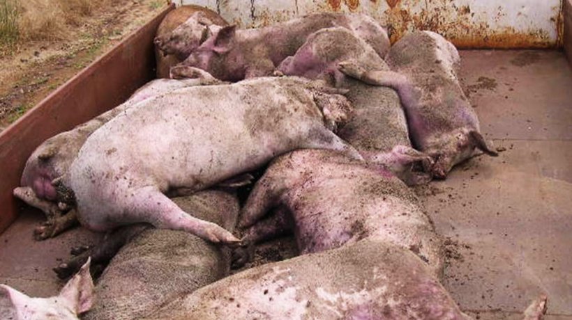
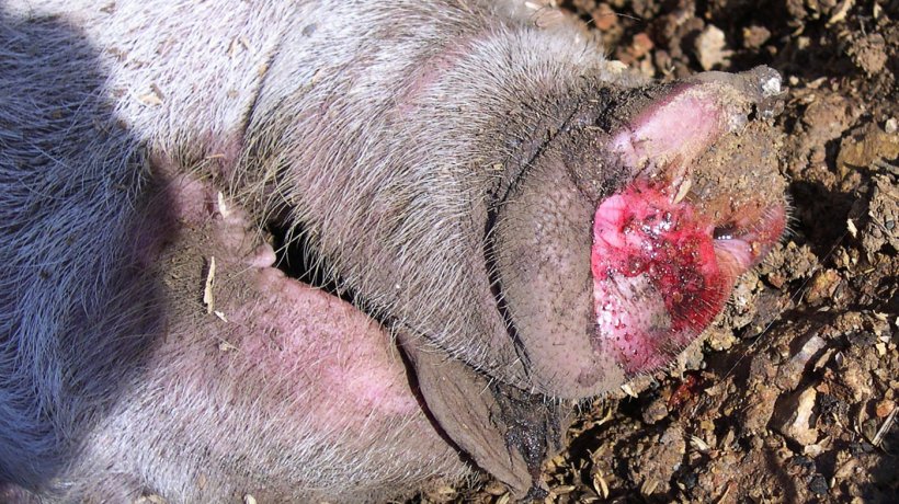
An expert team of veterinarians was directed to the affected sheds, where the dead and sick pigs were noted. The team confirmed clinically that the remaining pigs were generally dull and depressed and huddled together. These pigs often had a very high fever of 41ºC to 42ºC. The feet were normal. Many pigs had a discharge of froth from their nose, with bloody material in this froth in many cases. Some pigs had a bloody discharge from their anus. Many pigs had difficulty breathing and often lie on their sides, in an attempt to improve their breathing. Some pigs had a jaundiced yellow appearance to their skin and mucosae. Many pigs had spotty haemorrhages (petechiae) in the skin over their thorax and abdomen.
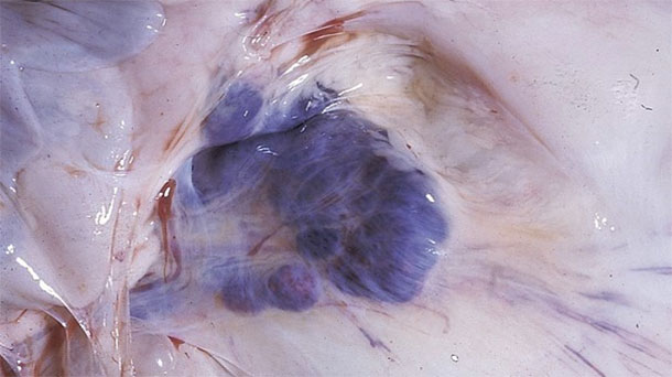
The team performed a full autopsy on several affected pigs. Care was taken to autopsy fresh acute cases. There were numerous splashy haemorrhages present in most of the body lymph nodes. For example, the mesenteric lymph nodes appeared like a string of large purple grapes.
The trachea and bronchi had marked amounts of frothy oedematous fluid, with some blood, confirming the source of the nasal discharge. The lungs had some diffuse congestion and oedema but were otherwise normal.
The liver and spleen were both markedly enlarged. The gall bladder had strong dark haemorrhages evident. The spleen enlargement was consistently massive, with a firm hard texture and 4 to 6 times normal size.
There was some ascites fluid present in the abdomen. The skin lesions of petechiae and jaundice were also noted at autopsy.

The farm workers placed in-feed and in-water antibiotics into the feed and water supply but without any discernible effect.
Differential diagnosis for outbreaks of high mortality in older pigs
The case study did not appear to be consistent with a toxic or physical problem such as an electrocution. Outbreaks of high mortality in groups of older pigs can be caused by several important infectious agents. The lung findings did not support a diagnosis of APP due to Actinobacillus pleuropneumoniae. Other possible causes of high mortality could include highly pathogenic PRRS and circovirus, with secondary infections, or other agents such as classical swine fever due to pestivirus. However, the findings of a severe haemorrhagic fever, with massive splenomegaly are highly suggestive of African swine fever (ASF).
The history, clinical signs and autopsy results suggest that a pathogenic strain of African swine fever (ASF) had colonised the farm in the previous weeks to the mortality outbreaks.
Further investigation and measures taken
The Dongbei region of China has direct land border connections with Russia, which has had numerous outbreaks of ASF due to the Georgia strain. The Russian outbreaks originated from farms close to the Black Sea ports, where shipping food waste had been fed to pigs. Analysis of the Chinese ASF strain has indicated that it is identical to the Georgia strain. It is therefore likely that the infection on this Dongbei farm came from trade and trucking routes into neighbouring farms and this case study farm.
The ASF virus is a large and complex DNA virus with multiple outer layers and numerous mechanisms aimed to avoid an immune reaction in the host pig.
There is therefore little likely value in development of a killed or subunit vaccine approach for ASF control. Strategies to develop a suitable live attenuated vaccine have not yet been successful.
The key to ASF control currently relies on depopulation of affected farms and prevention of new cases via strict biosecurity. ASF virus is mostly spread via contact with infected pigs (including wild boars) or pig materials, such as pork meat or offal products. There are few wild boars in the Dongbei area, however these are more common in southern Chinese areas such as the hill provinces.