Case history
In May 2014, two of three replacement gilts, which had died the night before, were brought to the Field Station for Epidemiology of the University of Veterinary Medicine Hanover for pathological examination. The gilts were part of a group of 25 replacement gilts of 180 days of age. The group had been born on a Danish farm and bred on a farm in Northern Germany. One week before the three gilts died, they had been delivered from this farm. During an examination by the herd attending veterinarian the unaffected gilts of this group had shown neither coughing or diarrhea nor other clinical signs of disease. Environmental reasons for the death of the three gilts had also been excluded by the herd vet.

Pathological findings
Both carcasses showed a good body condition and a pronounced cyanosis in the area of the ventral abdomen and the ventral throat. The skeletal muscles, especially the M. longissimus dorsi and the M. semimembranosus, showed pale areas, which were inelastic and easily torn (Figure 1). The myocardium of both gilts also possessed focal brightened alterations (Figure 2). In addition, there were multifocal subepicardial petechial hemorrhages. Furthermore, in one of the gilts a mild and focal, fibrinous pleurisy as well as fully encapsulated necrotic nodules in both diaphragmatic lobes of the lung were detected. The stomach of both gilts contained dry feed. Concerning the abdominal cavity, intestine, liver, spleen, kidney, urinary bladder, brain and bones no pathological lesions could be found.
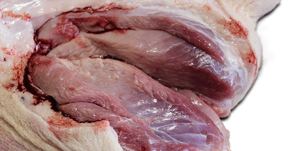
Figure 1: Pronounced paleness of the M. semimembranosus
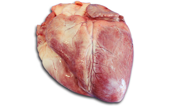
Figure 2: Focal brightened alterations of the myocardium
Sample collection
On the basis of these macroscopic findings, especially the alterations of the skeletal muscle the case was suspicious for vitamin E/selenium deficiency. Due to this fact, samples of the M. longissimus dorsi and the M. semimembranosus were fixed in formalin and sent to a laboratory (Lab 1) for histopathological examination. The encapsulated necrotic nodules detected in both diaphragmatic lobes of one gilt’s lung were assessed to be typical for a chronic infection with Actinobacillus pleuropneumoniae, which was probably not the cause of death. However, two samples of pulmonary tissue (altered and unaltered tissue) and a swab of the bronchial epithelium were collected for bacteriological investigation.
Recommendations for the herd veterinarian
According to the pathological findings the veterinarian of the Field station recommended to inject the remaining gilts with vitamin E and selenium.
Laboratory diagnoses
A. pleuropneumoniae was detected in the necrotic lung tissue and the bronchus via bacterial culture. The sample of lung tissue without pathological alterations was negative for bacterial pathogens. Concerning the formalin fixed samples of skeletal muscles and myocardium the histopathological investigation showed no microscopic lesions confirming the suspicion of a vitamin E/ selenium deficiency.
Interpretation of macroscopic and microscopic findings
When contacting the herd veterinarian again, we got the information that after the treatment with a vitamin E/ selenium emulsion no more gilts had died on the farm. For this reason and because of the impressive macroscopic findings typical for degenerative myopathies the suspicion of a vitamin E/ selenium deficiency was maintained. In order to be seen this confirmed, the formalin fixated samples of skeletal muscles and myocardium were sent to the Institute for Pathology and Forensic Veterinary Medicine of the University of Veterinary Medicine in Vienna for a second histopathological examination.
Final diagnoses
Repeating the histological investigation of the samples of skeletal muscles, the pathologist of the Institute for Pathology and Forensic Veterinary Medicine detected alterations in single muscle cells which represented different stages of myofiber degeneration. Some muscle fibers were swollen and a loss of striation was visible. Other parts of the muscles showed an evidence of acute myofiber necrosis (Figure 3). The microscopic findings of the myocardium were in compliance with an acute cardiac failure. These results confirmed the macroscopic findings characterized by paleness of muscles and inelastic, easily torn fibers, which aroused suspicion for a nutritional muscular degeneration mainly caused by vitamin E/ selenium deficiency.
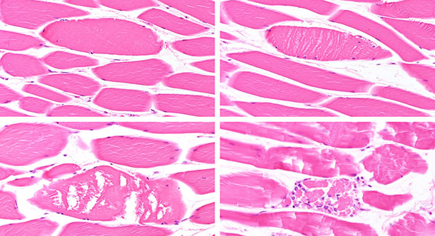
Figure 3: Different stages of myofiber degeneration/necrosis (from moderate swelling with loss of striation to complete desintegration and beginning resorption). Bar=80µm
Take-home messages
- Sudden death of gilts can be caused by vitamin E/selenium deficiency although vitamin E/selenium is usually supplemented in diets. Whether vitamin E/ selenium deficiency occurs in pigs beside those geographical areas already known for low levels of selenium in soil and plants (e. g. Scandinavian countries) needs to be investigated. Furthermore vitamin E can be destroyed by oxidation, if feed is stored for a longer period of time in unsuitable conditions.
- This case emphasizes that a suspicion based on clinical observations and macroscopic findings should be maintained even when it is not confirmed by the following in-depth investigations in the first trial.