

The first step to obtain a laboratory diagnosis of any clinical picture is proper sampling. This procedure is critical to achieve a successful analysis.
Regardless of the process to be analysed, the following premises must be followed when taking samples:

- Select representative animals: animals showing clinical signs, but at the early stages of the disease. This will reduce the presence of secondary or opportunistic agents, which can interfere with and even prevent detection of the primary cause of disease. Send samples of several animals (minimum 2-3). A single animal could not be representative of the problem at herd level.
- Select animals that have not been treated with antibiotics. In case of recurrent disease, an option is to leave some sentinel piglets with unmedicated feed. If this is not done, at least avoid the animals treated with antibiotics specific against the disease to be diagnosed.
- Perform necropsy and take samples immediately after death of the animal. Autolysis of samples makes diagnosis difficult, so euthanasia of representative animals is recommended. Animals dead for several hours must be avoided
Materials required for taking samples
- Latex gloves.
- Flasks with airtight closure. Whirl-pak bags or equivalent.
- Swabs with AMIES or STUART transport media. Transport media avoids desiccation of samples and maintains bacterial viability.
- Buffered formalin 4%: to be used for sample fixation to perform histopathology. Commercial formaldehyde is sold at 40%, therefore, to achieve the requested dilution, 9 parts of water must be mixed for each part of commercial formaldehyde.
- Styrofoam boxes and ice packs
SAMPLING:
Necropsies: whenever possible, organs must be submitted. In order to do that, the necropsy must be as complete as possible, ie, sending samples of all organs showing lesions, together with a detailed report.
Differential diagnosis of post-weaning diarrhoeas is crucial, so samples of all areas of the gastrointestinal tract where etiological agents are located must be taken (Table 1).
Table 1: Infectious agents causing diarrhoea in nursery-finishing pigs and target organs to be analysed
| Agent | Target organ |
| Enterotoxigenic Escherichia coli (ETEC) | Small intestine* |
| Clostridium perfringens | Small intestine |
| Brachyspira hyodysenteriae | Large intestine (colon and caecum) |
| Brachyspira pilosicoli | Large intestine (colon and caecum) |
| Lawsonia intracellularis | Ileum terminal, ileocaecal valve |
| Salmonella sp. | Small and large intestine |
| Virus: rotavirus, PEDV, TGE | Small intestine |
| Gastrointestinal parasites (nematodes). | Stomach, small and/or large intestine. |
* Clinical signs of post-weaning diarrhoea quite commonly occur together with signs of oedema disease, in which case the brain must be submitted too in order to assess lesions.
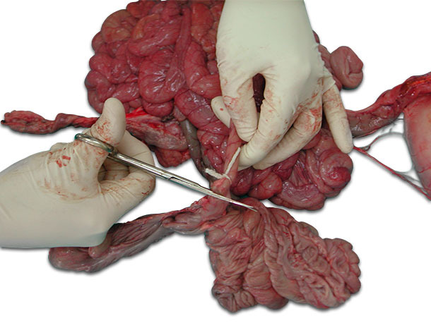
Sections of different portions of the intestine (jejunum, ileum and large intestine) must be sent, both fresh and fixed in formalin. Fresh samples will be used for microbiological study and other diagnostic techniques such as PCR. Before cutting the intestine sections, the ends of the intestine will be tied to avoid loss of content and contamination (photo 1). Each section will be placed separately into a hermetically sealed container or bag (photo 2). Content must be clearly identified.

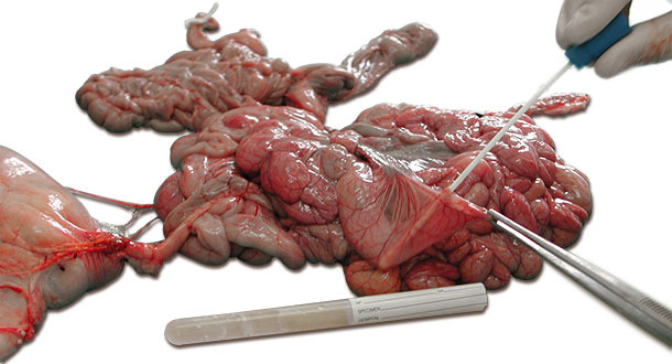
If submitting organs is not an option, swab samples can be taken directly from the intestinal sections to be analysed (photo 3).
The histopathological study of lesions is very important to make a complete diagnosis. It is recommended to send the samples already fixed in formalin, avoiding autolysis of organs during transport to the lab. Autolysis, which is especially critical in intestinal samples, prevents the histopathological diagnosis. Small sections of intestine will be sent in hermetic sealed flasks using a ratio of 1 part tissue to 10 parts of formalin.
Sampling live animals: if there are no suitable dead pigs and we want to avoid euthanasia of animals, it is possible to analyse faeces and rectal swabs.
Faecal samples should be collected directly from the rectal ampulla. To take the sample, introduce the swab and swirl it gently rubbing against the rectal walls.
Once the samples are taken they should be shipped to the laboratory as fast as possible. Transport with ice packs so samples are kept chilled (avoiding freezing) in a leak-proof container with enough protection to avoid damage in transit.


