

Influenza A virus or flu is a major cause of year-round acute respiratory disease in pigs of any age. In uncomplicated cases, the disease is mild and self-limiting, only lasting around seven days. The virulence of a virus is determined by the ability it has to invade tissues, the severity of the disease produced and the ability of the virus to spread within a susceptible population.
Endemic flu in pigs is predominantly caused by flu viruses of the subtypes H1N1, H1N2, and H3N2. Among these subtypes there are genetic variants that dominate within different regions throughout the world, often called genetic clusters or cluster types. Examples of these clusters would be the avian-like H1N1 viruses seen in Western Europe or the triple reassortant classic-like Alpha H1N1 viruses seen in Western Canada.

Within each of the genetic clusters, regardless of the subtype, there are virulent strains that cause more clinical disease (morbidity) and more deaths (mortality). While virulence is variable between viruses, the underlying mechanisms of infection remain consistent. Infection with flu viruses is initiated by the binding of hemagglutinin protein on the surface of the virus to sialic acid sugars on the surface of respiratory epithelial cells. These are the cells that line the nasal mucosa, trachea and airways of the lung (bronchi and bronchioles) – the smaller airways are targeted most.
Once the virus is internalized by the cell, it replicates and many virus particles are released causing cell death or necrosis of the cell. The necrotic cells slough off into the lumen of the airway and inflammatory cells (mostly neutrophils) respond. Within 24 to 48 hours the hallmark microscopic lesion of necropurulent bronchiolitis can be seen.
The remaining epithelial cells flatten out to cover the basement membrane (attenuation) and over the next 2-3 days the epithelial cells begin to multiply until they start to layer on top of each other (hyperplasia). Within 5-7 days, the virus has stopped replicating and the tissues begin to recover. The necrotic cells and inflammation are cleared out and the tissue can return to normal within 14-21 days of infection.
The gross lesions that you would see in the lung are polygonal areas representing lobules that are depressed (concave), dark red, and firm to the touch. These are areas where the air spaces (alveoli) surrounding the bronchioles have collapsed (lobular atelectasis) due to the obstruction caused by the necrotic cells and inflammatory cells within the airways (Figure 1b). This can be compared to normal (figure 1a).
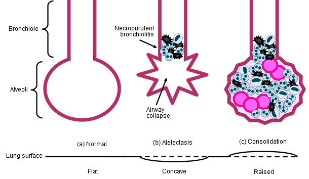
Figure 1. A A normal airway with the bronchiole leading into the lung lobule formed by several alveoli. B The bronchiole is filled with necrotic and inflammatory cells (necropurulent bronchiolitis) that are blocking air flow and causing collapse of the alveoli called atelectasis. C Secondary bacterial infection with Streptococcus suis, creating a consolidated lobule that is filled with bacteria, inflammatory cells, edema fluid and necrotic cells.
This is what happens in uncomplicated cases. Unfortunately, within the respiratory tract of a pig there are often pathogenic and opportunistic bacteria present. Common causes of secondary bacterial infections with flu are Streptococcus suis, Pasteurellamultocida, Haemophilus parasuis, Actinobacillussuis, and Bordetellabronchiseptica.
On a microscopic level, the air spaces fill with inflammatory cells responding to the bacterial colonies that are growing and protein rich fluid leaks out of the blood vessels (edema) along with fibrin. This is called consolidation. As the air spaces fill up so that the walls bend outward, we see the gross lesion of raised, dark red to purple, polygonal areas that are dense and meaty to the touch (Figure 1c).
The gross lesions for both uncomplicated flu infection and secondary bacterial infections are in the cranio-ventral parts of the lung because of the aerosol route of infection. With a pig standing on all four feet, the virus enters through the nose and descends down the trachea. When the virus reaches the lung, there are numerous short branches (bronchial tree) and gravitational pull that brings most of the virus into the small airways of the first part of the lung, the cranio-ventral portion. Most of the lesions are in the cranial and accessory lung lobes, but the lesions may also extend to the cranial-most portion of the caudal lung lobes.
In animals that have robust protection from their immune system provided by either a vaccine, previous infection or maternally-derived antibodies, the tissue damage caused by flu virus infection can be minimized. This happens when antibodies produced by the exposure are similar enough to the virus causing the infection. A perfect antigenic match is ideal because it will result in little to no virus replication/damage, shedding or clinical signs. However, if the virus is similar enough to have cross-reaction, the infection can be reduced to as few as 2-4 days.
