

The interaction between Mycoplasma hyopneumoniae and its host make M. hyopneumoniae one of the most important contributors to porcine respiratory disease. M. hyopneumoniae is transmitted via direct contact, most commonly in pigs older than 6 weeks; however, vertical transmission from sow/gilt to piglet also occurs. M. hyopneumoniae infection occurs when the bacteria attach to the ciliated epithelium lining the airway. Attachment leads to the clumping of cilia, ciliastasis, and death of affected ciliated epithelial cells. Damage to the ciliated epithelium results in a disruption of the mucociliary apparatus (disabling clearance of pathogens and dust particles in the airway) and expansion of the bronchus associated lymphoid tissue (BALT). These lesions contribute to the nonproductive cough seen with M. hyopneumoniae disease and the increased susceptibility to other respiratory tract pathogens. The time it takes between M. hyopneumoniae infection and appearance of clinical signs (most often when pigs are 7-12 weeks of age) is dependent on M. hyopneumoniae dose and strain, immune status, and management strategies.
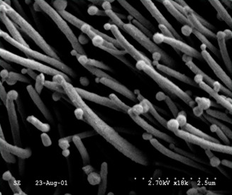

Image 1. Electron micrograph of Mycoplasma hyopneumoniae bacteria attached to the swine ciliated respiratory tract epithelium. Source: Dr. Carlos Pijoan.
Pneumonia can be seen as early as 1 week post M. hyopneumoniae infection, peaks 3-4 weeks post infection and the percentage of lung affected usually decreases by 5 weeks post infection. The time course of pneumonia coincides with the anti-M. hyopneumoniae immune response. Molecular patterns on the bacteria as well as signals from affected ciliated epithelial cells alert the immune system. Neutrophils infiltrate the peribronchiolar and perivascular areas of the lung within one week post infection and macrophages infiltrate within two weeks post infection. Both neutrophils and macrophages secrete pro-inflammatory cytokines, which in turn exacerbate the inflammatory reaction and attract lymphoid cells to the area of infection.
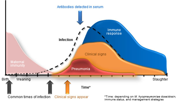
Figure 1. The dynamics of Mycoplasma hyopneumoniae infection, clinical signs, and host immune response over time. The different colored areas and arrows correspond with different components of M. hyopneumoniae infection and disease. All times are dependent upon M. hyopneumoniae dose/strain, immune status, and management strategies.
Lymphocytes are first found in affected areas of the lung about 2 weeks post infection. The presence of both T and B lymphocytes at the site of infection are important for M. hyopneumoniae clearance. T lymphocytes are vital in signaling B lymphocytes to produce IgA and IgG. IgA may neutralize the ability of M. hyopneumoniae to attach to ciliated epithelial cells; IgG is likely important in enhancing phagocytosis of bacteria. IgA and IgG can be found in the lung shortly after lymphocyte arrival; however, it may take up to 6 weeks for M. hyopneumoniae-specific antibodies to appear in the blood. T lymphocytes are also important in stimulating macrophages to be more efficient at killing bacteria they have phagocytosed. It has also been demonstrated that T lymphocytes in the blood respond to M. hyopneumoniae directly: they proliferate and produce IFNg when exposed to M. hyopneumoniae antigen in vitro.
M. hyopneumoniae also modulates the pig’s immune system. M. hyopneumoniae has been shown to have mitogenic properties and stimulate the production of pro-inflammatory cytokines. The presence of immune cells and their immunomodulatory proteins in the lung compounded by the pro-inflammatory milieu induced by the bacteria stimulates germinal center formation and expansion of the BALT. While a mature BALT may be more efficient at identifying and combating pathogens, the BALT may also serve as a reservoir of cells capable of being infected with pathogens, e.g., alveolar macrophages and PRRS. In well-managed herds M. hyopneumoniae may be relatively unimportant; however, in the presence of other respiratory pathogens, M. hyopneumoniae may set the stage for complex infections.
Antibiotic treatment often does not clear the infection, and while prevention via vaccines is beneficial to disease outcome especially in complex infection, vaccines do not prevent M. hyopneumoniae infection. Sow infections usually occur with the introduction of M. hyopneumoniae infected replacement animals, yet most sows will transmit the infection to only one litter of piglets as they will have cleared the infection and developed immunity to M. hyopneumoniae by their next litter. M. hyopneumoniae-specific immunity is transferred to piglets via colostrum/milk and has been shown to be at least partially-protective in those piglets. Maternally derived M. hyopneumoniae-specific immunity may contribute to the delay in onset of clinical signs in young pigs.
The economic impact of M. hyopneumoniae is largely influenced by management practices and barn conditions. Losses in daily gain may occur when 10-20% or more of lung is affected; however, herd performance may be inconsistently related to the degree of M. hyopneumoniae infection. Barns with high dust and poor ventilation, poor hygiene, or other animal stressors, may experience adverse effects to pig performance more rapidly.

