Both enzootic pneumonia, whose primary agent is Mycoplasma hyopneumoniae, and swine pleuropneumonia, caused by Actinobacillus pleuropneumoniae (APP), are respiratory diseases in pigs that are found worldwide and cause significant production and economic losses on pig farms, mainly during the growth and finishing phases.
Main lung lesions observed at the slaughterhouse
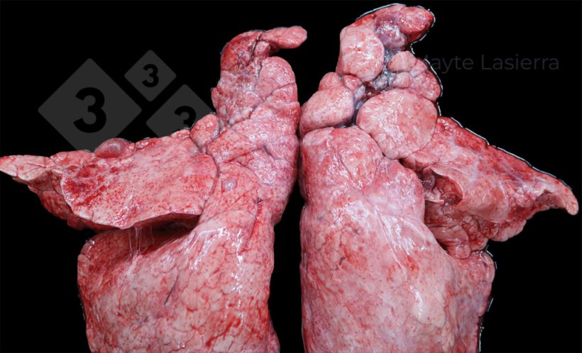
In lungs evaluated at the slaughterhouse, the most frequently observed lesions are cranio-ventral bronchopneumonia and pleuritis.

Cranio-ventral bronchopneumonia is mainly associated with enzootic pneumonia. These lesions appear as areas of consolidation, are hard to the touch, and range from gray to purple in color, depending on how old the lesion is and the secondary bacterial complications.

Bronchopneumonic process
Lesions appear approximately two weeks after infection and peak 2-4 weeks later.
Eight weeks after infection, bronchopulmonary lesions begin to recede, and scarring becomes apparent.

Scarring
Scarring may be complete 10-12 weeks after infection and may even disappear over time.
Analyzing the relationship between the prevalence of bronchopneumonic lungs and lungs with scars allows us to establish the approximate time during which the batch of pigs was affected by the lesions, thus estimating whether the lesions correspond to a:
- chronic process: began in the first half of finishing
- acute process: began during the second half of finishing
The presence of cranio-ventral bronchopneumonic lesions associated with Mycoplasma hyopneumoniae influences production indexes during the finishing period, causing varying degrees of loss in average daily gain, an increase in feed conversion ratio, and an increase in the finishing period, depending on the average surface area of the lungs affected by the lesions.
Pleuritis is frequently observed and consists of lesions affecting the dorsocaudal region of the lung, mainly associated with subacute or chronic forms of porcine pleuropneumonia (APP). It manifests as whitish areas on the visceral pleura of the diaphragmatic lobes, corresponding to fibrin deposits.
Cranial pleuritis

Cranial pleuritis

Cranial pleuritis

Dorsocaudal pleuritis
Pleuritis can also be observed affecting the cranial areas of the lung, creating adhesions between adjacent pulmonary lobes. These are usually associated with secondary respiratory processes of bacterial origin (Pasteurella multocida, Glaesserella parasuis, Streptococcus suis, etc.) and are called cranial pleuritis to differentiate them from those associated with APP.
How have lesions compatible with enzootic pneumonia evolved in Spain in recent years?
In Spain, after evaluating more than 6,500 batches of pigs and over a million lungs, we have observed a reduction in the degree of involvement by lesions compatible with Mycoplasma hyopneumoniae from 2016 to the present (Table 1 and Graph 1), both in:
- the prevalence of affected lungs (going from 49.96% to 23.71%)
- the average surface area of the lungs affected by lesions (going from 3.83% to 1.39%)
Table 1. Evolution of lung lesions from 2016 to 2024 in Spain.
| 2016 | 2017 | 2018 | 2019 | 2020 | 2021 | 2022 | 2023 | 2024 |
|---|---|---|---|---|---|---|---|---|
| Number of lungs evaluated | ||||||||
| 76,868 | 154,817 | 214,702 | 184,741 | 100,017 | 109,251 | 68,786 | 93,568 | 173,220 |
| % Bronchopneumonic lungs | ||||||||
| 49.96 | 42.64 | 41.50 | 37.60 | 32.34 | 31.11 | 35.56 | 28.65 | 24.33 |
| % Average lung area affected | ||||||||
| 3.83 | 3.16 | 2.79 | 2.53 | 2.12 | 2.37 | 2.29 | 1.96 | 1.45 |
| % Cranial pleurisy | ||||||||
| 13.24 | 13.90 | 21.36 | 21.15 | 21.30 | 18.28 | 10.11 | 10.08 | 12.67 |
| % Scarring | ||||||||
| 14.00 | 10.57 | 10.84 | 12.34 | 15.56 | 15.49 | 17.94 | 13.07 | 12.90 |
| % Dorsocaudal pleuritis | ||||||||
| 11.98 | 13.43 | 23.22 | 22.00 | 18.88 | 20.66 | 10.02 | 12.52 | 14.85 |
| APP rate | ||||||||
| 0.34 | 0.37 | 0.60 | 0.59 | 0.53 | 0.60 | 0.29 | 0.36 | 0.41 |

Graph 1. Evolution of lesions compatible with Mycoplasma hyopneumoniae.
We saw a slight increase during 2021 and 2022, possibly due to co-infections with highly virulent strains of PRRS that led to an increase in the severity of lesions observed and a general deterioration in farm health in certain areas.
This improvement observed over the years may be due both to better control strategies for the problems caused by Mycoplasma hyopneumoniae and to the increase in slaughter weight, and therefore in the age of the animals. This can be seen to some extent in the fact that the occurrence of scars does not decrease at the same rate as the prevalence of lesions, but rather seems to remain stable or even increase. Thus, it could be the case that many of the lesions have healed and are therefore not observed at slaughter as bronchopneumonic lesions, but as scars.
In animals systematically slaughtered at older ages, such as Iberian pigs or culled replacement sows, it is common to see a high number of scars without observing bronchopulmonary lesions.
What about pleuritis? How has it evolved?
With regard to dorso-caudal pleuritis associated with APP, there is no clear pattern of evolution, but rather there are fluctuations in the prevalence of lesions. It rose from 11.98% in 2016 to 23.22% in 2018, then fell back to previous levels in 2022, although there appears to be an increase in the prevalence of lesions again in the last two years evaluated (Table 1 and Graph 2).
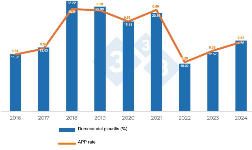
Graph 2. Evolution of lesions consistent with Actinobacillus pleuropneumoniae.
In the case of APP-associated lesions, the increase in lesion prevalence observed since 2018 could be associated with both the reduction in the widespread use of antibiotics during the nursery phase and the entry into the finishing phase, as well as the increase in average lactation length that occurred during those years. However, this variation in the prevalence and severity of lesions is less likely to follow a clear pattern, given the existence of different serotypes with varying degrees of virulence, whose prevalence also evolves over time. Nevertheless, both facts could be related, which would require studies associating the levels of lesion severity with the prevalence of different APP serotypes.
The value of farm-level analysis
While studying the evolution of lung lesions in a specific area or country can help us analyze the evolution of respiratory health in general, individualized studies on a farm or production pyramid are important because they allow us to estimate the degree to which these diseases affect production indexes, and sometimes it is the only way to monitor how the effectiveness of the measures implemented to control these diseases is evolving, especially in the case of Mycoplasma hyopneumoniae, given that production indexes can be affected by other factors not necessarily related to the disease.