

New technologies, such as computed tomography scan (CT scan), used as part of a genetic selection program, offer incredible opportunities to improve the selection of our boars. Thanks to this tool, we can collect information at large scale about the interior of our live pigs without compromising their welfare. These days, with so much talk about other technologies such as genomics, which undoubtedly play a key role in our improvement program, there is a phrase that we repeat like a mantra, reminding us of what is important: “In the age of genomics, phenotype is still King.” Despite the enormous contribution of genomics, collecting information on farms and test stations to evaluate the variable expression of an organism's genotype in a given environment (phenotype) remains fundamental. For this reason, the accuracy and variety of data collected have increased exponentially in recent years.
Computed tomography: technology applied to the selection of pigs of the present and the future
The use of computed tomography scan (CT) as an integral part of a selection program is the best example of how cutting-edge technology can contribute to selecting more robust and efficient pigs that are adapted to the industry's demands.

CT scan is a diagnostic imaging method widely used in human medicine and, to a lesser extent, in veterinary medicine. It is based on the use of X-rays to scan the entire animal, taking cross-sectional views (1,100 images for an entire pig) that allow a complete three-dimensional image of the animal to be created, including all its structures, bones, muscles, and organs.
What are the advantages of computed tomography as part of a genetic improvement program?
- Analysis of carcass composition (yield of cuts and lean percentage) directely in the live boars that are candidates to be selected, allowing for more accurate estimation of genetic values and the development of new genetic values (e.g., for ham yield or fat coverage in ham). Specific selection of boars for the needs of each producer.
- It allows for a significant increase in the number of animals analyzed compared to other tools, such as carcass analysis at slaughterhouses. Thousands of animals are tested each year.
- Incorporation of phenotypes that cannot be evaluated in any other way in live animals, such as the presence of osteochondrosis or arthritis lesions in the joints that can lead to lameness in later stages of the animal's life.
- New phenotypes that allow for the selection of more robust animals based on other characteristics (cardiac, hepatic, pulmonary volume, etc.).
How was CT incorporated into the genetic selection program?
It was first used in 2008 as an alternative way of analyzing the carcass composition of boars undergoing testing, which could replace the slaughterhouse dissection protocols carried out on related animals that was used previously.
For years, image analysis was performed manually, identifying different anatomical areas and tissue types (bone, fat, and muscle), which was already a significant advance. In 2016, a more automated method of CT image analysis was implemented, allowing different major cuts (ham, shoulder, loin, and belly) to be distinguished. The emergence of artificial intelligence (AI) technologies was key to automating this process, which was essential for scaling up this tool, increasing the number of animals analyzed in a more efficiente way.
However, AI requires a prior training process, which necessitates a huge database of previously analyzed images to serve as models. In our case, this database was available thanks to years of previous CT analysis, during which significant progress had already been made in tissue segmentation and the identification of voxels (the smallest units that make up a three-dimensional image, similar to pixels in a photograph) belonging to different tissues.
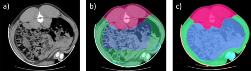
Figure 1. CT images, a) without segmentation, b) segmentation prior to AI implementation, c) with manual corrections of the segments used during the AI training process. The pink area corresponds to the spine, blue to the intestines, and green to the abdominal wall, with the limb segment appearing in turquoise.
The quality of this AI-based automation process can be evaluated through genetic analyses and by estimating the heritability of the traits calculated based on these images. The higher the heritability, the lower the noise due to other factors, and the more reliable the information obtained and used in calculating genetic values. Heritability in the lean percentage and lean composition of the different cuts is moderate to high (range 0.33 to 0.75), demonstrating the value of this technology.
The automation of the analysis process and the standardization of the procedure, as well as the boar management protocols, allow more than 15,000 boars (from four genetic lines, both dam and sire lines) to be analyzed each year with this technology.
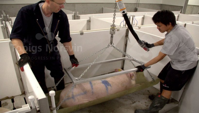
Figure 2. The animals are sedated to keep them calm during the process.
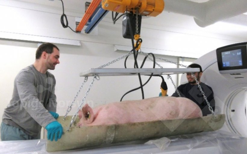
Figure 3. With an articulated stretcher system, animals are moved from the preparation area to the CT scanner.
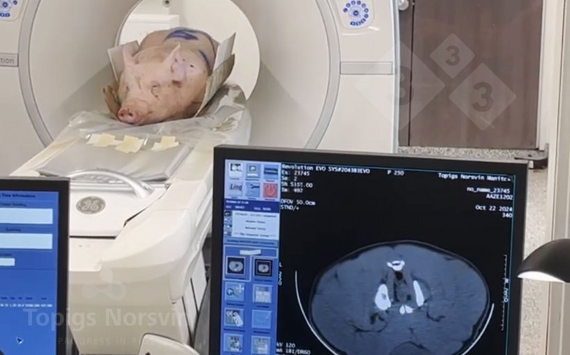
Figure 4. The boars are positioned prone on the CT scanner. The entire process takes less than 10 minutes.
How does CT help us improve the robustness of pigs?
Being able to see the inside of our live pigs in detail opens the door to evaluating aspects that we had not even considered until now. The possibilities are endless, and we are only beginning to understand and take advantage of them. Currently, many efforts are focused on identifying qualities in animals at an early age that can be associated with more robust and long-lived animals. The following are currently being evaluated:
The presence of lesions in the head of the femur and humerus to identify arthritis or osteochondrosis processes. This assessment is part of the selection objective for all genetic lines. The evaluation process includes a manual component that will soon be replaced by an automated process for segmenting and quantifying lesions, which will also allow for the evaluation of more bone surfaces.
The size and shape of the scapula have been evaluated and correlated with sow longevity and the presence of ulcers and other lesions on the shoulder of adult sows.
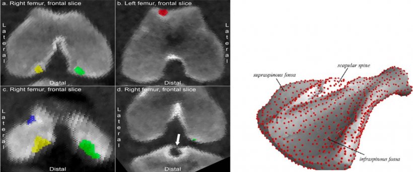
Figure 5. Right: Frontal and lateral views of femoral heads with lesions on the articular surface. Left: Three-dimensional image of the scapula.
Other phenotypes that could be of interest: Research is currently underway to develop new phenotypes related to pig viscera, such as the volume of organs, including the heart and lungs, which could be related to the robustness of the animals and their tolerance to stress. However, the automation of these phenotypes poses additional challenges, as these organs behave dynamically, changing shape and size with breathing and heartbeat, which complicates the automatic analysis process. Research is currently being conducted in cooperation with the Faculty of Veterinary Medicine at NMBU University (Norway) to integrate CT information with information collected using electrocardiography (ECG).
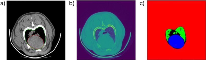
Figure 6. CT images showing cardiac volume and profile (a,b) and automatic differentiation of different tissue types (c).
Computed tomography is an excellent example of how cutting-edge technology can be used as a fundamental part of a pig selection program, allowing us to learn details about our pigs that we could not have even imagined years ago. Because “in the age of genomics, phenotype is still King.”