

Laboratory diagnostics: Ileitis (Lawsonia intracellularis)
What laboratory diagnostic methods can I use to diagnose ileitis? Which one should I choose according to the situation? How do I interpret the results?
The pig sector events all around the world
Pig health: news and articles on PRRS, PCV2, biosecurity, etc, Pig disease guide, atlas of pathology, clinical cases…
Biocheck.UGent is an independent, risk-based, scientific scoring system for assessing the quality of your on-farm biosecurity.
A visual and practical step-by-step guide on how to perform a necropsy on a pig.
All the information about ASF: how to recognize the disease, how it is transmitted, pictures of lesions, latest news, guides, etc.
Description of the most important diseases and conditions in pigs
Images of major swine diseases
Pig disease diagnostic tool
Definition for the most commonly used pig terms
Simulator that calculates the amount of drug to add to the water when using a flow dispenser.
Pig Prices by countries. Pork production and trade. News of the pig market and the raw materials
The latest slaughter pig prices in the most important pig markets. Check the evolution of the historical prices in charts and in several currencies.
Latest quotations for the main commodities used in pig feed. Historical graphs with the pig price and estimated feed price.
Figures & trends in pig numbers, pork production and pork trade.
Global production and trade data for the most important raw materials
Articles on nutrition and pig feeding, characteristics of raw materials and additives for pig feed. Prices of raw materials
Latest quotations for the main commodities used in pig feed. Historical graphs with the pig price and estimated feed price.
Technical sheets of the main raw materials and additives used in swine feed. They include a comparison of nutritional values from various sources, product
Global production and trade data for the most important raw materials
Definition for the most commonly used pig terms
Use this tool to diagnose problems with the feed conversion ratio. Click on the flowchart or on the buttons within the text to navigate through the different parts of the tool.
Articles on genetics and pig reproduction: genetic improvement, genomics, artificial insemination, use of hormones
Compare production data, calculate the number of sow, nursery, and finishing spaces, and visualize your tasks on the work schedule by type of BMS.
Tool that allows you to calculate the replacement rate in your farm
Definition for the most commonly used pig terms
Use this tool to find out why your farrowing rate is less than ideal. Click on the flowchart or on the buttons found within the text to navigate through the different parts of the tool.
Management, pig farm management, work planning in each production stage: management in gestation, grow finish, batch farrowing
Compare production data, calculate the number of sow, nursery, and finishing spaces, and visualize your tasks on the work schedule by type of BMS.
Tool that allows you to calculate the replacement rate in your farm
Definition for the most commonly used pig terms
Design of facilities and equipment for pig farms: building design, climate control, feeding systems, etc.
Definition for the most commonly used pig terms
Use this tool to explore which slurry management strategy best fits your situation. Click on the flow chart or on the buttons within the text to navigate through the different parts of the tool.
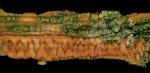
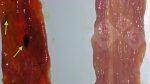
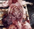
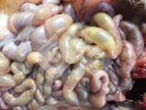
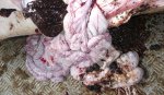
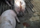
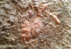
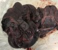
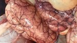
The disease has three different presentations: porcine intestinal adenopathy (PIA), an abnormal proliferation of the intestinal mucosa; necrotic enteritis (NI), where the proliferated cells of the small intestine die and get detached with a gross thickening of the small intestine (hosepipe gut); and acute hemorrhagic ileitis,an hyper-acute inflammation which causes massive bleeding.
Alternative names: Lawsonia intracellularis, porcine intestinal adenopathy (PIA), necrotic enteritis (NI), hosepipe gut
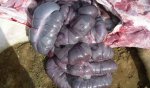
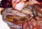
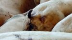
Describes a group of conditions that imply pathological changes in the small intestine associated to the bacteria Lawsonia intracellularis, which, as its name indicates, lives inside the intestinal cells. The organism is present in all (or almost all) farms. The disease has three different presentations: porcine intestinal adenopathy (PIA), an abnormal proliferation of the intestinal mucosa; necrotic enteritis (NI), where the proliferated cells of the small intestine die and get detached with a gross thickening of the small intestine (hosepipe gut); and acute hemorrhagic ileitis, an hyper-acute inflammation which causes massive bleeding. In the latter there is massive bleeding into the small intestine, hence the name of bloody gut and this is the most common form in growing pigs and gilts.
The organism is impossible to keep out of farms probably because it also infects other species including horses. Infected feces are the major vehicle for spread around the farm.
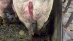
Gilts
Usually showing the hemorrhagic acute disease:.
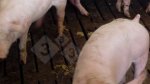
Weaners and growers
PIA and NI clinical signs are different form hemorrhagic acute ileitis.
Porcine Intestinal Adenopathy (PIA)
Lactating piglets
Not fully understood.
Based on the clinical picture, post-mortem examinations, histology of the gut wall and demonstrating the presence of the organism in feces by PCR test. A serological ELISA test is also available.
Use antibiotics to treat sick pigs.
Strategic use of antibiotics at high risk times.
A good hygiene and the use of oral or injectable vaccines are recommended.
What laboratory diagnostic methods can I use to diagnose ileitis? Which one should I choose according to the situation? How do I interpret the results?

